PET/MRI has shown neuroreceptors in the brains of obese people appear to respond differently to food cues, a German group noted on 27 June at the Society for Nuclear Medicine and Molecular Imaging annual meeting.
The finding adds to our basic understanding of the mechanisms underlying obesity and reveals a potential new target for treatments, said Dr. Swen Hesse of the University of Leipzig.
"The search for interventions to achieve sustainable weight loss reaches high priority including thorough investigation of biological and behavioral mechanisms in individuals with obesity," Hesse said.
Obesity is considered a global epidemic and poses a major challenge for healthcare systems worldwide, with more than 1 billion people considered obese. The risks of heart attack, stroke, and type 2 diabetes are significantly increased in people with obesity.
In this study, Hesse and colleagues investigated receptors in the brain's cholinergic system, which plays a significant role in the brain's reward system. Specifically, they targeted a type of nicotinic acetylcholine receptor called alpha-4 beta-2 nicotinic (a4b2), which among other functions is involved in how people decide which foods are most desirable, Hesse explained.
"We aimed to measure changes in a4b2 nicotinic acetylcholine receptors (nAChR) found in the cholinergic system in response to high-caloric food cues," he said.
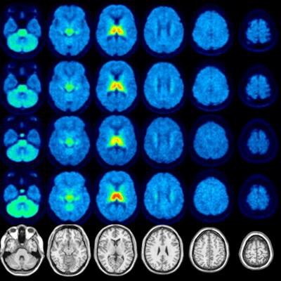
The group enrolled 15 people with obesity (average body mass index [BMI], 38 +/- 3 kg/m2) and 16 normal-weight people for PET/MRI scans with F-18 flubatine, a radiotracer developed more than 20 years ago to bind to a4b2 receptors. Participants underwent hybrid PET/MRI twice on separate days, once while in a resting state and once under "food cue stimulation" -- the subjects were shown food pictures just prior to the scans, Hesse said.
Distribution volumes of a4b2 nicotinic acetylcholine receptors in the brain of normal-weight volunteers and volunteers with obesity. Image courtesy of Swen Hesse, MD.In the resting state, no significant difference in total distribution volume of F-18 flubatine was noted between the groups. However, while viewing photos of food, the total distribution volumes (VT) of F-18 flubatine was higher in obese subjects in the brain's thalamus region where a4b2 receptors are located compared to the control group (28.8 ± 2.1 vs. 25.1 ± 1.9, p = 0.03), according to the findings.
"These changes are associated with an attentional bias toward incentive food cues and likely changes in sensory perception," Hesse noted.
Further research is warranted, Hesse said, Ultimately, the research shows that PET/MRI can reveal novel pathways in the brain's reward center in people with obesity, with the findings representing a model of "higher distractibility" towards rewarding food cues, he concluded.


![Examples of ultrasound findings and techniques. (A) Images in a 39-year-old male patient with a mass in the left thigh. The mass is heterogeneous on the B-mode US image (compared with the patient in D) and showed increased microvascularity (superb microvascular imaging [SMI]) and shear-wave elastography (SWE) values. Undifferentiated pleomorphic sarcoma was diagnosed at biopsy (with pleomorphic rhabdomyosarcoma in surgical specimen). (B) Images in an 18-year-old male patient with a mass in the left leg. The mass is hypoechoic on the B-mode image, with no other findings suggestive of malignancy. The lesion is in contact with the cortex of the tibia, which is slightly irregular. CT revealed a doubtful anteromedial tibial erosion. The microvascular study demonstrated high vascularization, suggestive of malignancy. Periosteal Ewing sarcoma was diagnosed with both histologic and immunohistochemical confirmation. (C) Images in a 69-year-old female patient with a lump growing on the outside of the left leg. Multiple SWE examinations were performed (please note the high values obtained in the measurements, whereas the color map highlights the stiffness relative to adjacent tissues). SMI showed areas of increased vascularization to target for sampling. Undifferentiated spindle cell sarcoma was diagnosed at biopsy, with residual leiomyosarcoma in the surgical specimen after neoadjuvant therapy. (D) Images in a 56-year-old female patient with a mass in the right thigh. The mass is heterogeneous at both B-mode ultrasound (similar to patient A) and MRI (coronal T2-weighted spectral attenuated inversion recovery [SPAIR]; T1-weighted pre-contrast and postcontrast imaging), which even shows uptake after the administration of paramagnetic contrast material, which is traditionally suggestive of malignancy. Low values at SMI and elastography are suggestive of benignity. Spindle cell lipoma was diagnosed at biopsy, with atypical spindle cell lipomatous tumor in the surgical specimen.](https://img.auntminnieeurope.com/mindful/smg/workspaces/default/uploads/2026/08/images-radiol250278fig2.APCFLSvX6p.jpg?auto=format%2Ccompress&fit=crop&h=100&q=70&w=100)


![Examples of ultrasound findings and techniques. (A) Images in a 39-year-old male patient with a mass in the left thigh. The mass is heterogeneous on the B-mode US image (compared with the patient in D) and showed increased microvascularity (superb microvascular imaging [SMI]) and shear-wave elastography (SWE) values. Undifferentiated pleomorphic sarcoma was diagnosed at biopsy (with pleomorphic rhabdomyosarcoma in surgical specimen). (B) Images in an 18-year-old male patient with a mass in the left leg. The mass is hypoechoic on the B-mode image, with no other findings suggestive of malignancy. The lesion is in contact with the cortex of the tibia, which is slightly irregular. CT revealed a doubtful anteromedial tibial erosion. The microvascular study demonstrated high vascularization, suggestive of malignancy. Periosteal Ewing sarcoma was diagnosed with both histologic and immunohistochemical confirmation. (C) Images in a 69-year-old female patient with a lump growing on the outside of the left leg. Multiple SWE examinations were performed (please note the high values obtained in the measurements, whereas the color map highlights the stiffness relative to adjacent tissues). SMI showed areas of increased vascularization to target for sampling. Undifferentiated spindle cell sarcoma was diagnosed at biopsy, with residual leiomyosarcoma in the surgical specimen after neoadjuvant therapy. (D) Images in a 56-year-old female patient with a mass in the right thigh. The mass is heterogeneous at both B-mode ultrasound (similar to patient A) and MRI (coronal T2-weighted spectral attenuated inversion recovery [SPAIR]; T1-weighted pre-contrast and postcontrast imaging), which even shows uptake after the administration of paramagnetic contrast material, which is traditionally suggestive of malignancy. Low values at SMI and elastography are suggestive of benignity. Spindle cell lipoma was diagnosed at biopsy, with atypical spindle cell lipomatous tumor in the surgical specimen.](https://img.auntminnieeurope.com/mindful/smg/workspaces/default/uploads/2026/08/images-radiol250278fig2.APCFLSvX6p.jpg?auto=format%2Ccompress&fit=crop&h=112&q=70&w=112)