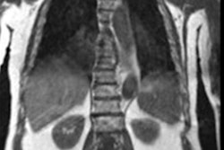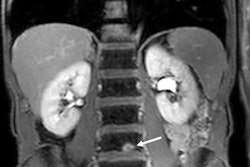
Whole-body MRI with diffusion-weighted imaging (DWI) is a viable alternative to F-18 FDG-PET/CT imaging for lung cancer staging, according to research presented at the recent ECR meeting in Vienna.
"Whole-body MRI is an ideal radiation-free imaging tool for the staging of lung cancer with good diagnostic accuracy," said presenter Dr. Ajith Antony of All India Institute of Medical Sciences (AIIMS) in New Delhi, India.
Lung cancer is the second most commonly diagnosed cancer around the world, and the leading cause of cancer-related death, Antony noted. The current gold standard for staging the disease is either contrast-enhanced CT of the chest and upper abdomen or F-18 FDG PET/CT imaging. But both of these exams expose patients to high doses of radiation.
Whole-body MRI shows promise as an attractive alternative for lung cancer staging because it does not impart radiation, but there have been few studies evaluating its performance for this indication, Antony said. To compare the two exams, he and colleagues conducted a study that included 31 patients with biopsy-proven lung cancer.
The whole-body MRI protocol consisted of the following sequences:
- Axial fluid attenuation inversion recovery (FLAIR)
- Axial short tau inversion recovery (STIR)
- Axial dual fast field echo (FFE) T1-weighted
- Axial diffusion-weighted imaging
- Contrast enhanced axial T1-weighted mDIXON (fat suppression technique)
The F-18 FDG PET/CT protocol consisted of patient fasting for four hours before radiopharmaceutical injection; a whole-body scan taken about an hour after injection, with a low-dose CT scan first followed by a PET scan. Images were reconstructed using iterative techniques.
Antony and colleagues found that whole-body MRI -- particularly when used with diffusion-weighted imaging (DWI) -- showed high performance across a variety of measures for staging different levels of lung cancer.
| Overall staging with whole body MRI | |||
| Measure | Stage I and II | Stage III | Stage IV |
| Sensitivity | |||
| With DWI | 100% | 93.3% | 100% |
| Without DWI | 96.6% | 80% | 100% |
| Specificity | |||
| With DWI | 100% | 100% | 94.1% |
| Without DWI | 100% | 100% | 88.2% |
| AUC | |||
| With DWI | 1 | 0.97 | 0.98 |
| Without DWI | 0.98 | 0.90 | 0.94 |
Antony conceded that one of the study's limitations was its small sample size and that pathological confirmation of suspicious lung nodes wasn't available. Yet the study results remain promising, he said.
"Whole-body [MRI] can be used for [lung cancer] staging with comparable accuracy to the present gold standard," he concluded.


![Examples of ultrasound findings and techniques. (A) Images in a 39-year-old male patient with a mass in the left thigh. The mass is heterogeneous on the B-mode US image (compared with the patient in D) and showed increased microvascularity (superb microvascular imaging [SMI]) and shear-wave elastography (SWE) values. Undifferentiated pleomorphic sarcoma was diagnosed at biopsy (with pleomorphic rhabdomyosarcoma in surgical specimen). (B) Images in an 18-year-old male patient with a mass in the left leg. The mass is hypoechoic on the B-mode image, with no other findings suggestive of malignancy. The lesion is in contact with the cortex of the tibia, which is slightly irregular. CT revealed a doubtful anteromedial tibial erosion. The microvascular study demonstrated high vascularization, suggestive of malignancy. Periosteal Ewing sarcoma was diagnosed with both histologic and immunohistochemical confirmation. (C) Images in a 69-year-old female patient with a lump growing on the outside of the left leg. Multiple SWE examinations were performed (please note the high values obtained in the measurements, whereas the color map highlights the stiffness relative to adjacent tissues). SMI showed areas of increased vascularization to target for sampling. Undifferentiated spindle cell sarcoma was diagnosed at biopsy, with residual leiomyosarcoma in the surgical specimen after neoadjuvant therapy. (D) Images in a 56-year-old female patient with a mass in the right thigh. The mass is heterogeneous at both B-mode ultrasound (similar to patient A) and MRI (coronal T2-weighted spectral attenuated inversion recovery [SPAIR]; T1-weighted pre-contrast and postcontrast imaging), which even shows uptake after the administration of paramagnetic contrast material, which is traditionally suggestive of malignancy. Low values at SMI and elastography are suggestive of benignity. Spindle cell lipoma was diagnosed at biopsy, with atypical spindle cell lipomatous tumor in the surgical specimen.](https://img.auntminnieeurope.com/mindful/smg/workspaces/default/uploads/2026/08/images-radiol250278fig2.APCFLSvX6p.jpg?auto=format%2Ccompress&fit=crop&h=100&q=70&w=100)


![Examples of ultrasound findings and techniques. (A) Images in a 39-year-old male patient with a mass in the left thigh. The mass is heterogeneous on the B-mode US image (compared with the patient in D) and showed increased microvascularity (superb microvascular imaging [SMI]) and shear-wave elastography (SWE) values. Undifferentiated pleomorphic sarcoma was diagnosed at biopsy (with pleomorphic rhabdomyosarcoma in surgical specimen). (B) Images in an 18-year-old male patient with a mass in the left leg. The mass is hypoechoic on the B-mode image, with no other findings suggestive of malignancy. The lesion is in contact with the cortex of the tibia, which is slightly irregular. CT revealed a doubtful anteromedial tibial erosion. The microvascular study demonstrated high vascularization, suggestive of malignancy. Periosteal Ewing sarcoma was diagnosed with both histologic and immunohistochemical confirmation. (C) Images in a 69-year-old female patient with a lump growing on the outside of the left leg. Multiple SWE examinations were performed (please note the high values obtained in the measurements, whereas the color map highlights the stiffness relative to adjacent tissues). SMI showed areas of increased vascularization to target for sampling. Undifferentiated spindle cell sarcoma was diagnosed at biopsy, with residual leiomyosarcoma in the surgical specimen after neoadjuvant therapy. (D) Images in a 56-year-old female patient with a mass in the right thigh. The mass is heterogeneous at both B-mode ultrasound (similar to patient A) and MRI (coronal T2-weighted spectral attenuated inversion recovery [SPAIR]; T1-weighted pre-contrast and postcontrast imaging), which even shows uptake after the administration of paramagnetic contrast material, which is traditionally suggestive of malignancy. Low values at SMI and elastography are suggestive of benignity. Spindle cell lipoma was diagnosed at biopsy, with atypical spindle cell lipomatous tumor in the surgical specimen.](https://img.auntminnieeurope.com/mindful/smg/workspaces/default/uploads/2026/08/images-radiol250278fig2.APCFLSvX6p.jpg?auto=format%2Ccompress&fit=crop&h=112&q=70&w=112)