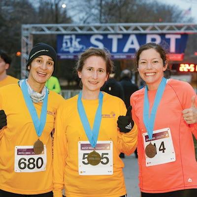
Sports-minded RSNA attendees had a bonanza on day 3 of the congress. A 5k fun run took place in the morning, and then four World Cup soccer games were broadcast live in a dedicated space in McCormick Place.
Early on a brisk Tuesday morning in Chicago, a total of 441 runners (out of the anticipated 31,000 in-person congress attendees) completed the RSNA 5k fun run. The event was definitely one for early birds, starting at 6.30 a.m. in Arvey Field, South Grant Park.
Advance registration for the event cost $40 per runner, while the onsite entry fee was $50. "The funds will help improve patient care by supporting research and education in radiology through grants and awards to individuals and institutions that advance radiologic research, education, and practice," noted a report in Wednesday's RSNA Daily Bulletin.
Top finishers in the women's division were (left to right) Graciela Garrido (silver), Guggenberger Konstanze (gold), and Emily Ambinder (bronze). Courtesy of RSNA Daily Bulletin.
In the men's division, the medals went to (left to right) Erik Fredenberg (bronze), Francesco Mungai (gold), and Romain Labas (silver). Courtesy of RSNA Daily Bulletin.Any runners who were feeling weary after their exertions could put their feet up and watch live action from the soccer World Cup.
The host country, Qatar, United Arab Emirates, is nine hours ahead of Chicago, so most of the matches are being played during the working day in the U.S. This prompted the congress organizers to set aside an area for attendees to watch the action on a big screen.
RSNA delegates take a break between sessions to enjoy some action from the World Cup in Qatar, UAE.In Tuesday's fixtures, Senegal beat Ecuador 2-1, Netherlands beat Qatar 2-0, USA beat Iran 1-0, and England beat Wales 3-0. Netherlands will meet the U.S. on Saturday in the last 16/knockout phase of the tournament, and Senegal will face England on Sunday.
The World Cup final takes place on Sunday, 18 December at 3 p.m. local time.


![Examples of ultrasound findings and techniques. (A) Images in a 39-year-old male patient with a mass in the left thigh. The mass is heterogeneous on the B-mode US image (compared with the patient in D) and showed increased microvascularity (superb microvascular imaging [SMI]) and shear-wave elastography (SWE) values. Undifferentiated pleomorphic sarcoma was diagnosed at biopsy (with pleomorphic rhabdomyosarcoma in surgical specimen). (B) Images in an 18-year-old male patient with a mass in the left leg. The mass is hypoechoic on the B-mode image, with no other findings suggestive of malignancy. The lesion is in contact with the cortex of the tibia, which is slightly irregular. CT revealed a doubtful anteromedial tibial erosion. The microvascular study demonstrated high vascularization, suggestive of malignancy. Periosteal Ewing sarcoma was diagnosed with both histologic and immunohistochemical confirmation. (C) Images in a 69-year-old female patient with a lump growing on the outside of the left leg. Multiple SWE examinations were performed (please note the high values obtained in the measurements, whereas the color map highlights the stiffness relative to adjacent tissues). SMI showed areas of increased vascularization to target for sampling. Undifferentiated spindle cell sarcoma was diagnosed at biopsy, with residual leiomyosarcoma in the surgical specimen after neoadjuvant therapy. (D) Images in a 56-year-old female patient with a mass in the right thigh. The mass is heterogeneous at both B-mode ultrasound (similar to patient A) and MRI (coronal T2-weighted spectral attenuated inversion recovery [SPAIR]; T1-weighted pre-contrast and postcontrast imaging), which even shows uptake after the administration of paramagnetic contrast material, which is traditionally suggestive of malignancy. Low values at SMI and elastography are suggestive of benignity. Spindle cell lipoma was diagnosed at biopsy, with atypical spindle cell lipomatous tumor in the surgical specimen.](https://img.auntminnieeurope.com/mindful/smg/workspaces/default/uploads/2026/08/images-radiol250278fig2.APCFLSvX6p.jpg?auto=format%2Ccompress&fit=crop&h=100&q=70&w=100)


![Examples of ultrasound findings and techniques. (A) Images in a 39-year-old male patient with a mass in the left thigh. The mass is heterogeneous on the B-mode US image (compared with the patient in D) and showed increased microvascularity (superb microvascular imaging [SMI]) and shear-wave elastography (SWE) values. Undifferentiated pleomorphic sarcoma was diagnosed at biopsy (with pleomorphic rhabdomyosarcoma in surgical specimen). (B) Images in an 18-year-old male patient with a mass in the left leg. The mass is hypoechoic on the B-mode image, with no other findings suggestive of malignancy. The lesion is in contact with the cortex of the tibia, which is slightly irregular. CT revealed a doubtful anteromedial tibial erosion. The microvascular study demonstrated high vascularization, suggestive of malignancy. Periosteal Ewing sarcoma was diagnosed with both histologic and immunohistochemical confirmation. (C) Images in a 69-year-old female patient with a lump growing on the outside of the left leg. Multiple SWE examinations were performed (please note the high values obtained in the measurements, whereas the color map highlights the stiffness relative to adjacent tissues). SMI showed areas of increased vascularization to target for sampling. Undifferentiated spindle cell sarcoma was diagnosed at biopsy, with residual leiomyosarcoma in the surgical specimen after neoadjuvant therapy. (D) Images in a 56-year-old female patient with a mass in the right thigh. The mass is heterogeneous at both B-mode ultrasound (similar to patient A) and MRI (coronal T2-weighted spectral attenuated inversion recovery [SPAIR]; T1-weighted pre-contrast and postcontrast imaging), which even shows uptake after the administration of paramagnetic contrast material, which is traditionally suggestive of malignancy. Low values at SMI and elastography are suggestive of benignity. Spindle cell lipoma was diagnosed at biopsy, with atypical spindle cell lipomatous tumor in the surgical specimen.](https://img.auntminnieeurope.com/mindful/smg/workspaces/default/uploads/2026/08/images-radiol250278fig2.APCFLSvX6p.jpg?auto=format%2Ccompress&fit=crop&h=112&q=70&w=112)