
Dr. Dolores Ferrer Puchol of the Hospital Universitario de la Ribera de Alzira in Valencia, Spain, has received a top award at the 36th Congress of the Spanish Society of Medical Radiology (SERAM) and XXXI CIR.
Puchol received the award for most cited article for "Selective intra-arterial embolization as a treatment for hemorrhoidal pathology," published in the journal Radiologia.
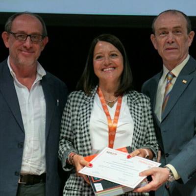
Dr. Dolores Ferrer Puchol (center) receives the SERAM award for most cited article. Image courtesy of the SERAM.In the article, Puchol and colleagues describe how they used right femoral artery or radial artery access to catheterize the inferior mesenteric artery, proceeding to the superior rectal artery with a 2.7 F microcatheter to catheterize and embolize each distal branch distally with polyvinyl alcohol particles (300-500 µm) and proximally with coils (2-3 mm). Patients were discharged 24 hours after the procedure and clinically followed up at one month by anoscopy.
In 20 patients, the authors achieved technical success in 18 (90%) patients and clinical success in 15 (83.4%).
During the awards ceremony, the author thanked SERAM for its efforts to contribute to the science of radiology in Spain and for awarding prizes that encourage its members to continue working for science.


![Overview of the study design. (A) The fully automated deep learning framework was developed to estimate body composition (BC) (defined as subcutaneous adipose tissue [SAT] in liters; visceral adipose tissue [VAT] in liters; skeletal muscle [SM] in liters; SM fat fraction [SMFF] as a percentage; and intramuscular adipose tissue [IMAT] in deciliters) from MRI. The fully automated framework comprised one model (model 1) to quantify different BC measures (SAT, VAT, SM, SMFF, and IMAT) as three-dimensional (3D) measures from whole-body MRI scans. The second model (model 2) was trained to identify standardized anatomic landmarks along the craniocaudal body axis (z coordinate field), which allowed for subdividing the whole-body measures into different subregions typically examined on clinical routine MRI scans (chest, abdomen, and pelvis). (B) BC was quantified from whole-body MRI in over 66,000 individuals from two large population-based cohort studies, the UK Biobank (UKB) (36,317 individuals) and the German National Cohort (NAKO) (30,291 individuals). Bar graphs show age distribution by sex and cohort. BMI = body mass index. (C) After the performance assessment of the fully automated framework, the change in BC measures, distributions, and profiles across age decades were investigated. Age-, sex-, and height-adjusted body composition reference curves were calculated and made publicly available in a web-based z-score calculator (https://circ-ml.github.io).](https://img.auntminnieeurope.com/mindful/smg/workspaces/default/uploads/2026/05/body-comp.XgAjTfPj1W.jpg?auto=format%2Ccompress&fit=crop&h=112&q=70&w=112)


