Radiologists in Belgium are remembering the renowned interventional radiologist Prof. Julien Struyven, who died in Uccle, Brussels, on 21 April at the age of 84.
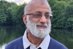
 Prof. Julien Struyven. All photos courtesy of Dr. Christian Delcour.
Struyven was the first person in Belgium to perform several important interventional procedures. In 1969, he performed the first coronary angiography, and he performed the first Belgian embolization in 1974, the first esophageal dilatation in 1979, and the first coronary dilatation in 1980.
"He is without doubt an icon in radiology," wrote Prof. Erik Ranschaert, PhD, a radiologist at St. Nikolaus Hospital, Eupen, and visiting professor at Ghent University, in his tribute to Struyven posted on the Radiologen BeNe Facebook page on 26 April.
His international career was brilliant, Dr. Christian Delcour, chef de service at Le Centre Hospitalier Universitaire de Charleroi -- ISPPC, Brussels, noted in a personal tribute to his mentor.
Contribution to CIRSE
Struyven was born on 14 November 1937 in Ougrée, located in the province of Liège.
He made a significant contribution to the development of the Cardiovascular and Interventional Radiological Society of Europe (CIRSE), having helped to create the organization through the merger of two separate societies: the European College of Angiography and the European Society of Cardiovascular Radiology.
In 1990, he organized CIRSE's annual congress in Brussels, and he served as its president between 1994 and 1997.
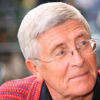
 Struyven (in center of photo, with glasses) was a valued and active member of the Belgian community of radiologists, particularly during the 1980s and 1990s. Other leading figures in this photo include Prof. Robert Dondelinger (with brown tie and jacket and moustache) and Prof. Danielle Balériaux.
"On his impulse, CIRSE developed itself in a very impressive way, with an exponential increase in the number of its members," said Delcour. "It also got organized in an exemplary way, becoming a representative European body in the field of interventional radiology and building a very high-level European teaching structure."
Delcour was a radiology trainee at the Erasmus Hospital of the Free Brussels University (FBU) under Struyven's supervision and chairmanship between 1980 and 1984.
Substantial legacy
In 1984, Struyven was appointed professor of radiology at the FBU. He authored and co-authored more than 250 articles listed in Medline, as well as numerous books and chapters.
For many years, he also played a central role within the Belgian Society of Radiology (BSR), acting as both chairman and secretary general for several years. Until very recently, he was an active member of the Executive Committee and was in charge of the website.
It is due in part to his inspiration and guidance that the BSR now has such a thriving interventional section.
 Struyven was an active supporter of radiology in developing countries, particularly Africa. Dr. Roger Manono Katoto, from the Democratic Republic of the Congo, worked as a radiologist in Brussels during the 1980s.
In addition to Struyven's numerous academic merits, he was very endearing, profoundly human, and loved to serve others, Delcour wrote. He is also remembered for his good sense of humor and as a knowledgeable modern art lover. He also traveled extensively, especially in Africa.
A farewell ceremony for Julien Struyven, followed by a cremation, will occur at Uccle crematorium on 28 April at 9.30 a.m. He is survived by his sons Pierre-Emmanuel and Jean-Frédéric, and sister Emmy, and he was the widower of his beloved wife Dr. Juliette Aderca.
"Art is the answer" was the simple message at the top of the announcement message for the ceremony.