A one-day training course can yield significant and sustained performance improvement for radiologists who are interpreting CT colonography (CTC) exams, according to research published online on 15 February in Radiology.
This new paper provides extra information from the authors following their initial article published on 9 June 2020 in Clinical Radiology.
In a large prospective multicenter trial performed in the U.K., a group of radiologists who attended a one-day, individualized workshop on CTC reporting had a marked increase in sensitivity for detecting clinically relevant colorectal neoplasia than a control group of radiologists who did not receive the training.
What's more, these sensitivity improvements were still evident at six and 12 months after the training, and they were "independent of career experience, lesion location, or morphological characteristics," wrote a research team led by Dr. Anu Obaro of University College London.
Most jurisdictions don't require testing, accreditation, or performance monitoring for reporting of CTC exams, despite its status as the first-choice radiologic test for colorectal cancer screening and examination of symptomatic patients, according to the researchers.
"This contrasts with processes for colonoscopy and other cancer screening programs," they wrote.
In an attempt to address this deficiency, the researchers performed a prospective, multicenter cluster-randomized trial including 69 hospitals and 139 radiologists from England and Wales. All radiologists in the study were experienced in CTC interpretations and reported these exams routinely. Of the participants, 80 were randomized into an intervention group that received the one-day CTC training course with personalized feedback. The remaining 59 radiologists did not receive any additional training.
The researchers sought to assess whether the training could improve the radiologists' diagnostic sensitivity for clinically significant lesions (≥ 6 mm or higher) and if so, to determine the durability of these improvements. They evaluated the participating radiologists' performance in interpreting 10 CTC scans at time points of one, six, and 12 months later.
| Impact of 1-day CTC training course on radiologist performance | ||
| Radiologists with no extra training | Radiologists who attended 1-day CTC training with individualized feedback | |
| Baseline | 43.4% | 49.4% |
| 1 month | 42.4% | 66.4% |
| 6 months | 50.5% | 66.4% |
| 12 months | 44.4% | 63.7% |
| Detection of flat lesions | 28.5% | 55% |
With the exception of the baseline performance, all differences were statistically significant. In other findings, the researchers did not observe an association between the positive effect of training with prior CTC career experience.
The researchers noted that training and ongoing assessment should be mandated for practitioners interpreting CTC exams, and certainly within national screening programs.
"Such accreditation is already stipulated for breast cancer screening and would align CTC with colonoscopy screening," the authors wrote. "Our data suggest that radiologists would welcome this, and previous surveys have found that radiologists favor accreditation and assessment."
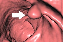
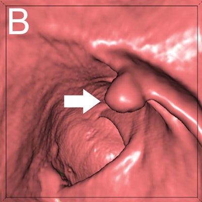
Editor's note: the image used to introduce this article on the homepage is a CTC of a 9-mm polyp (arrow) in the descending colon of a 78-year-old woman. Virtual fly-through 3D reconstructions were used for exact polyp localization. Courtesy of Dr. Sergio Grosu and colleagues from Ludwig Maximilian University of Munich and the RSNA.

![Examples of ultrasound findings and techniques. (A) Images in a 39-year-old male patient with a mass in the left thigh. The mass is heterogeneous on the B-mode US image (compared with the patient in D) and showed increased microvascularity (superb microvascular imaging [SMI]) and shear-wave elastography (SWE) values. Undifferentiated pleomorphic sarcoma was diagnosed at biopsy (with pleomorphic rhabdomyosarcoma in surgical specimen). (B) Images in an 18-year-old male patient with a mass in the left leg. The mass is hypoechoic on the B-mode image, with no other findings suggestive of malignancy. The lesion is in contact with the cortex of the tibia, which is slightly irregular. CT revealed a doubtful anteromedial tibial erosion. The microvascular study demonstrated high vascularization, suggestive of malignancy. Periosteal Ewing sarcoma was diagnosed with both histologic and immunohistochemical confirmation. (C) Images in a 69-year-old female patient with a lump growing on the outside of the left leg. Multiple SWE examinations were performed (please note the high values obtained in the measurements, whereas the color map highlights the stiffness relative to adjacent tissues). SMI showed areas of increased vascularization to target for sampling. Undifferentiated spindle cell sarcoma was diagnosed at biopsy, with residual leiomyosarcoma in the surgical specimen after neoadjuvant therapy. (D) Images in a 56-year-old female patient with a mass in the right thigh. The mass is heterogeneous at both B-mode ultrasound (similar to patient A) and MRI (coronal T2-weighted spectral attenuated inversion recovery [SPAIR]; T1-weighted pre-contrast and postcontrast imaging), which even shows uptake after the administration of paramagnetic contrast material, which is traditionally suggestive of malignancy. Low values at SMI and elastography are suggestive of benignity. Spindle cell lipoma was diagnosed at biopsy, with atypical spindle cell lipomatous tumor in the surgical specimen.](https://img.auntminnieeurope.com/mindful/smg/workspaces/default/uploads/2026/08/images-radiol250278fig2.APCFLSvX6p.jpg?auto=format%2Ccompress&fit=crop&h=100&q=70&w=100)


![Examples of ultrasound findings and techniques. (A) Images in a 39-year-old male patient with a mass in the left thigh. The mass is heterogeneous on the B-mode US image (compared with the patient in D) and showed increased microvascularity (superb microvascular imaging [SMI]) and shear-wave elastography (SWE) values. Undifferentiated pleomorphic sarcoma was diagnosed at biopsy (with pleomorphic rhabdomyosarcoma in surgical specimen). (B) Images in an 18-year-old male patient with a mass in the left leg. The mass is hypoechoic on the B-mode image, with no other findings suggestive of malignancy. The lesion is in contact with the cortex of the tibia, which is slightly irregular. CT revealed a doubtful anteromedial tibial erosion. The microvascular study demonstrated high vascularization, suggestive of malignancy. Periosteal Ewing sarcoma was diagnosed with both histologic and immunohistochemical confirmation. (C) Images in a 69-year-old female patient with a lump growing on the outside of the left leg. Multiple SWE examinations were performed (please note the high values obtained in the measurements, whereas the color map highlights the stiffness relative to adjacent tissues). SMI showed areas of increased vascularization to target for sampling. Undifferentiated spindle cell sarcoma was diagnosed at biopsy, with residual leiomyosarcoma in the surgical specimen after neoadjuvant therapy. (D) Images in a 56-year-old female patient with a mass in the right thigh. The mass is heterogeneous at both B-mode ultrasound (similar to patient A) and MRI (coronal T2-weighted spectral attenuated inversion recovery [SPAIR]; T1-weighted pre-contrast and postcontrast imaging), which even shows uptake after the administration of paramagnetic contrast material, which is traditionally suggestive of malignancy. Low values at SMI and elastography are suggestive of benignity. Spindle cell lipoma was diagnosed at biopsy, with atypical spindle cell lipomatous tumor in the surgical specimen.](https://img.auntminnieeurope.com/mindful/smg/workspaces/default/uploads/2026/08/images-radiol250278fig2.APCFLSvX6p.jpg?auto=format%2Ccompress&fit=crop&h=112&q=70&w=112)