Ultrasound images used for statistical shape modeling can help predict hip dysplasia in infants and whether abduction treatment devices are needed, according to Dutch research published on 25 January in Radiology.
A team led by Dr. Joshua Bonsel from University Medical Center Utrecht in the Netherlands found that modeling ultrasound images of well-centered stable dysplastic hips in infants predicted which hips developed to normal or remained dysplastic, as well as determined which hips benefited from the Pavlik harness treatment.
"Statistical shape modeling identified acetabular shape characteristics on ultrasound images of Graf type II hips that predict acetabular development and show a correlation with treatment," Bonsel and colleagues wrote. "The statistical shape modeling was also able to differentiate acetabular shape differences due to probe positioning from true anatomic variations."
Developmental hip dysplasia is common in newborns, with previous research suggesting an incidence between 1.5 and 20 per 1,000 births. While patients with Graf types III and IV are referred for immediate treatment, there is no consensus on how and when to treat well-centered stable infant hips with acetabular dysplasia. These hips have a bony roof angle between 43° and 59°, whereas a value of 60° or more is considered normal.
The researchers wrote that many well-centered stable hips are currently treated with an abduction device despite the possibility that they could develop into normal hips, leading to possible overtreatment.
"Current diagnostic parameters for stable developmental dysplasia of the hip only poorly correlate with outcome and cannot be used to identify hips that will have persistent or residual hip dysplasia with or without treatment," the study authors wrote.
Statistical shape modeling can quantify the shape of the acetabulum on ultrasound images. Bonsel et al said using ultrasound images can render more data for development of the hip than angle measurements alone.
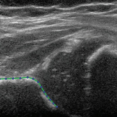
 Hip ultrasound image in a 3-month-old girl at baseline. No contrast material was used. The presented hip is outlined with the acetabular shape model, consisting of 13 points. The researchers wrote that through statistical shape modeling from ultrasound images, hip dysplasia, and treatment can be better predicted. Image courtesy of RSNA.
They wanted to identify acetabular shape characteristics of Graf type II hips and identify which hips benefited from Pavlik harness treatment. They also wanted to identify artifacts due to probe positioning.
"In the context that around 80% of Graf II hips will develop to normal without treatment, there is a need to extract more data from ultrasound images to identify hips that are at risk for persistent or residual hip dysplasia at follow-up," Bonsel and colleagues wrote.
The team looked at data from baseline ultrasound images in 97 infants with an average age of 3.37 years. Out of these, 89 were girls and the other eight were boys. The group found 90 cases of Graf type IIb hip dysplasia, 52 of which were treated with Pavlik harness.
The modeling created several shape modes from the 13 landmarks placed on ultrasound images of each hip. These modes showed shape variations of hip shapes on imaging sets.
The researchers found that shape modes 2 and 3 of the modeling were significantly linked to persistent hip dysplasia on ultrasound images, with odds ratios of 0.43 and 2.39, respectively. Mode 2 was also linked to residual hip dysplasia on pelvic radiographs, with an odds ratio of 0.09 (p = 0.002).
Pavlik harness treatment was found to be beneficial in patients with negative mode 2 values, with an odds ratio of 12.46 (p = 0.01).
Bonsel and colleagues called for more studies to validate the predictive value of the identified shape modes. However, they added that an acetabular shape mode may lead to more accurate prognosis and prevent overtreatment, which could have important medical and socioeconomic benefits.
"Also, statistical shape modeling in software might help the examiner identify the ideal projection plane in a series of ultrasound images in real time," they wrote.