Dear AuntMinnieEurope Member,
Why wasn't disgraced radiologist Dr. Anthony Carmine Donadio erased from the U.K. medical register earlier? How rigorous were the checks made on his credentials and work over 20 years? How many patients suffered as a result of his bad practice and dishonesty? Did he work in Italy after he completed his training in Naples in 1992? And is he still working in Ukraine, where he is believed to be living now?
These are some of the unanswered questions resulting from the case of Donadio. All in all, it's frightening to contemplate the cost of the legal action taken against him since 2017, but thankfully, he has now been publicly shamed and prevented from working again, at least in the U.K.
In other news, the findings of an important study of pulmonary embolism (PE) were published this week. The researchers analyzed the management and outcome of over 1,400 patients who presented to 18 emergency departments in France and Spain between October 2019 and June 2020 with PE symptoms.
Further evidence of the progress being made by medical imaging in Turkey was provided at RSNA 2021. Musculoskeletal specialists from Ankara collected one of the 24 hotly contested magna cum laude awards in Chicago for their work on the challenges, optimization, and applications of zero echo-time imaging. Don't miss our news report on their groundbreaking efforts. Find out more in the MRI Community.
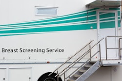
In another Turkish presentation at the RSNA meeting, investigators from Istanbul showed how middle-income countries can benefit from the use of artificial intelligence for breast screening to combat workforce shortages. Learn more in the Women's Imaging Community.
Finally, when it comes to radiology training, the Scandinavians tend to be among the most innovative and creative people in the world, so it's not surprising to see that Norway has implemented a novel system. Interestingly, the new curriculum covers the insecurities of both patients and doctors.