Using dual-energy CT (DECT), researchers have diagnosed bone disease in the fossilized jaw of a Tyrannosaurus rex named Tristan Otto, according to research presented this week at RSNA 2021. The work shows how DECT can be a noninvasive tool for paleontologists to investigate fossils.
"We hypothesized that DECT could potentially allow for quantitative noninvasive element-based material decomposition and thereby help paleontologists in characterizing unique fossils," said Dr. Charlie Hamm, a radiologist at Charité University Hospital in Berlin, who collaborated on the research with scientists from the Museum of Natural Science in Berlin, Chicago's Field Museum, and the University of Illinois at Chicago.
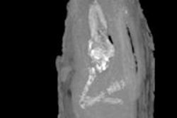
Otto's left lower jaw was successfully analyzed for bone disease using a clinical CT scanner with DECT. This technique allowed the researchers to scan the portion of the jaw called the left dentary, which in Otto's case measures a little over 2.5 ft (79.5 cm) long and 3 inches (81 mm) wide.
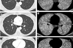
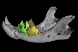
 CT reconstructions of the tooth-bearing part of the left dentary. (A) Reconstruction of the conventional CT images in lateral view showing well-preserved anatomical structures, such as the replacement teeth. The arrow indicates the focal exophytic mass -- the abnormal growth that sticks out from the surface of the tissue -- on the ventral surface at the level of the third to fifth tooth roots. (B) The DECT-based calcium material map shows a homogeneous mineral distribution, while (C) the fluorine material map shows significant mineral accumulation in the center of the exophytic mass and adjacent tooth roots (arrowhead). All images courtesy of Hamm et al.
To overcome the challenges of the fossil's high density, the researchers manipulated the CT scanner's current and voltage to minimize artifacts and improve image quality.
The left dentary of the jaw showed a diffuse thickening of nearly the entire dentary and a mass on its surface that extended to the root of one of the teeth. Inside the mass, DECT detected a significant accumulation of the element fluorine, which is associated with decreased bone density. The mass and fluorine accumulation findings supported a diagnosis of tumefactive osteomyelitis, an infection of the bone.
"The noninvasive density- and element-based material decomposition of fossilized bone revealed that fluorine could serve as an imaging biomarker for areas with decreased bone density, helping paleontologists to investigate fossils without the need to harm their integrity," the authors wrote.
Tristan Otto is one of the most complete T. rex skeletons ever found. The fossilized skeleton dates back approximately 68 million years to the Late Cretaceous period, when T. rex roamed widely across the western U.S. The commercial paleontologist who found the skeleton in Montana in 2010 sold it to an investment banker who dubbed it "Tristan Otto" before loaning it out to the Museum of Natural Science in Berlin in Germany.
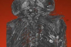
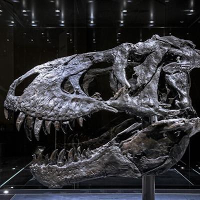
 The "Tristan Otto" T-rex skull that was examined by researchers.
The successful fossil imaging of Tristan Otto is likely just the beginning of a new role for radiology in paleontological research, according to the authors.
"While this is a proof-of-concept study, noninvasive DECT imaging that provides structural and molecular information on unique fossil objects has the potential to address an unmet need in paleontology, avoiding defragmentation or destruction," Hamm said.
Going forward, the DECT approach may prove useful in other paleontological applications, such as confirming the age of fossils and differentiating actual bone from replicas.
"The experimental design, including the use of a clinical CT scanner, will allow for broad applications," suggested Oliver Hampe, PhD, senior scientist and vertebrate paleontologist from the Museum of Natural Science in Berlin.