MRI shows that mild to moderate COVID-19 during pregnancy does not have any harmful effects on fetal brains, according to research being presented on Wednesday, 1 December, at the RSNA annual meeting.
A team led by Dr. Sophia Stöcklein from the Ludwig Maximilian University of Munich in Germany found normal fetal brain development, including cortical folding and brain stem segmentation, in their study of fetuses whose expectant mothers had COVID-19.
"While evidence regarding vertical transmission of SARS-CoV-2 during pregnancy remains sparse, our study did not reveal signs of infection or impaired development of the fetal brain on fetal MRI," Stöcklein and colleagues wrote.
It's been two years since the COVID-19 pandemic began, and evidence has shown that pregnant women are more vulnerable to the SARS-CoV-2 virus that causes COVID-19. What that means for developing fetuses, though, is up for debate, including whether or not the virus can be passed on from mother to fetus.
Stöcklein et al wanted to use fetal MRI to find out whether fetal brain development is affected in cases of SARS-CoV-2 infection during pregnancy. They looked at data from 33 patients with SARS-CoV-2 infection during pregnancy.
The women were on average 28.4 weeks into their pregnancies, with COVID-19 symptoms beginning on average 18.3 weeks into the pregnancy. The most common symptoms experienced by the women were loss or a reduced sense of smell and taste, dry cough, fever, and shortness of breath.
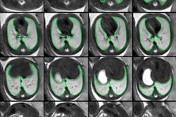
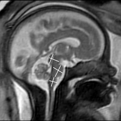
A research team led by Dr. Sophia Stöcklein from the Ludwig Maximilian University of Munich in Germany used MRI to show that fetal brains had age-appropriate developments even as their expectant mothers presented with mild to moderate COVID-19 symptoms. Image courtesy of the RSNA.The team found that brain development characteristics, such as opercularization of the Sylvian fissure, cortical folding, and transverse cerebellar diameter were age-appropriate in all fetuses. They also found that the diameter and craniocaudal extent of the pons, medulla oblongata, midbrain, and cerebellar vermis were all within age-appropriate limits.
"No calcifications, edema, or ventricular enlargement were detected. Seven fetuses showed mild asymmetry of the lateral ventricles with a maximum difference of 4 mm," they wrote.
However, Stöcklein said patients presented with mild or moderate symptoms and did not need hospitalization. She also said vaccination should be considered for pregnant women.
The team will be following the patients for the next five years to assess their neurological developments.


![Examples of ultrasound findings and techniques. (A) Images in a 39-year-old male patient with a mass in the left thigh. The mass is heterogeneous on the B-mode US image (compared with the patient in D) and showed increased microvascularity (superb microvascular imaging [SMI]) and shear-wave elastography (SWE) values. Undifferentiated pleomorphic sarcoma was diagnosed at biopsy (with pleomorphic rhabdomyosarcoma in surgical specimen). (B) Images in an 18-year-old male patient with a mass in the left leg. The mass is hypoechoic on the B-mode image, with no other findings suggestive of malignancy. The lesion is in contact with the cortex of the tibia, which is slightly irregular. CT revealed a doubtful anteromedial tibial erosion. The microvascular study demonstrated high vascularization, suggestive of malignancy. Periosteal Ewing sarcoma was diagnosed with both histologic and immunohistochemical confirmation. (C) Images in a 69-year-old female patient with a lump growing on the outside of the left leg. Multiple SWE examinations were performed (please note the high values obtained in the measurements, whereas the color map highlights the stiffness relative to adjacent tissues). SMI showed areas of increased vascularization to target for sampling. Undifferentiated spindle cell sarcoma was diagnosed at biopsy, with residual leiomyosarcoma in the surgical specimen after neoadjuvant therapy. (D) Images in a 56-year-old female patient with a mass in the right thigh. The mass is heterogeneous at both B-mode ultrasound (similar to patient A) and MRI (coronal T2-weighted spectral attenuated inversion recovery [SPAIR]; T1-weighted pre-contrast and postcontrast imaging), which even shows uptake after the administration of paramagnetic contrast material, which is traditionally suggestive of malignancy. Low values at SMI and elastography are suggestive of benignity. Spindle cell lipoma was diagnosed at biopsy, with atypical spindle cell lipomatous tumor in the surgical specimen.](https://img.auntminnieeurope.com/mindful/smg/workspaces/default/uploads/2026/08/images-radiol250278fig2.APCFLSvX6p.jpg?auto=format%2Ccompress&fit=crop&h=100&q=70&w=100)


![Examples of ultrasound findings and techniques. (A) Images in a 39-year-old male patient with a mass in the left thigh. The mass is heterogeneous on the B-mode US image (compared with the patient in D) and showed increased microvascularity (superb microvascular imaging [SMI]) and shear-wave elastography (SWE) values. Undifferentiated pleomorphic sarcoma was diagnosed at biopsy (with pleomorphic rhabdomyosarcoma in surgical specimen). (B) Images in an 18-year-old male patient with a mass in the left leg. The mass is hypoechoic on the B-mode image, with no other findings suggestive of malignancy. The lesion is in contact with the cortex of the tibia, which is slightly irregular. CT revealed a doubtful anteromedial tibial erosion. The microvascular study demonstrated high vascularization, suggestive of malignancy. Periosteal Ewing sarcoma was diagnosed with both histologic and immunohistochemical confirmation. (C) Images in a 69-year-old female patient with a lump growing on the outside of the left leg. Multiple SWE examinations were performed (please note the high values obtained in the measurements, whereas the color map highlights the stiffness relative to adjacent tissues). SMI showed areas of increased vascularization to target for sampling. Undifferentiated spindle cell sarcoma was diagnosed at biopsy, with residual leiomyosarcoma in the surgical specimen after neoadjuvant therapy. (D) Images in a 56-year-old female patient with a mass in the right thigh. The mass is heterogeneous at both B-mode ultrasound (similar to patient A) and MRI (coronal T2-weighted spectral attenuated inversion recovery [SPAIR]; T1-weighted pre-contrast and postcontrast imaging), which even shows uptake after the administration of paramagnetic contrast material, which is traditionally suggestive of malignancy. Low values at SMI and elastography are suggestive of benignity. Spindle cell lipoma was diagnosed at biopsy, with atypical spindle cell lipomatous tumor in the surgical specimen.](https://img.auntminnieeurope.com/mindful/smg/workspaces/default/uploads/2026/08/images-radiol250278fig2.APCFLSvX6p.jpg?auto=format%2Ccompress&fit=crop&h=112&q=70&w=112)