Dear AuntMinnieEurope Member,
If anybody knows what radiology services are really like in England, that person must be Dr. Giles Maskell.
Over an 18-month period before the pandemic, his team visited 143 hospitals and clinics. He presented his verdict in a powerful session held at the UK Imaging & Oncology Congress, which draws to a close today. Maskell did not hold back, and his honest assessment deserves close scrutiny. Find out more in the MRI Community.
In many ways, New Zealand has seemed like a shining example of how to minimize the disruption and distress caused by COVID-19, but the kiwis do occasionally get it wrong. A damning report published this week exposed the bad practice of a midwife who didn't bother to read radiology reports. She said she was too busy. Go to the Women's Imaging Community for our full coverage.
How can lung ultrasound be used effectively in suspected cases of COVID-19? Dr. Dirk-André Clevert and his colleagues in the European Society of Radiology's Ultrasound Subcommittee have addressed this question in a new consensus statement.
In other news, researchers from Turkey have found that an artificial intelligence algorithm was able to identify kidney stones -- even very small stones -- at an extremely high level of accuracy on CT.
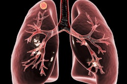
Chest CT isn't usually associated with breast cancer detection, but maybe it should be: The modality can identify incidental, suspicious breast lesions in women undergoing the exam for other reasons, and many of these lesions are malignant, according to a German study.


![Overview of the study design. (A) The fully automated deep learning framework was developed to estimate body composition (BC) (defined as subcutaneous adipose tissue [SAT] in liters; visceral adipose tissue [VAT] in liters; skeletal muscle [SM] in liters; SM fat fraction [SMFF] as a percentage; and intramuscular adipose tissue [IMAT] in deciliters) from MRI. The fully automated framework comprised one model (model 1) to quantify different BC measures (SAT, VAT, SM, SMFF, and IMAT) as three-dimensional (3D) measures from whole-body MRI scans. The second model (model 2) was trained to identify standardized anatomic landmarks along the craniocaudal body axis (z coordinate field), which allowed for subdividing the whole-body measures into different subregions typically examined on clinical routine MRI scans (chest, abdomen, and pelvis). (B) BC was quantified from whole-body MRI in over 66,000 individuals from two large population-based cohort studies, the UK Biobank (UKB) (36,317 individuals) and the German National Cohort (NAKO) (30,291 individuals). Bar graphs show age distribution by sex and cohort. BMI = body mass index. (C) After the performance assessment of the fully automated framework, the change in BC measures, distributions, and profiles across age decades were investigated. Age-, sex-, and height-adjusted body composition reference curves were calculated and made publicly available in a web-based z-score calculator (https://circ-ml.github.io).](https://img.auntminnieeurope.com/mindful/smg/workspaces/default/uploads/2026/05/body-comp.XgAjTfPj1W.jpg?auto=format%2Ccompress&fit=crop&h=112&q=70&w=112)


