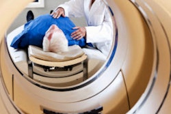
A PET/MRI scanner has been installed at the Misr Radiology Center in Cairo, Egypt, making it the first facility in Africa to have a PET/MRI system, according to a report posted by the European Society for Hybrid, Molecular, and Translational Imaging (ESHIMT) .
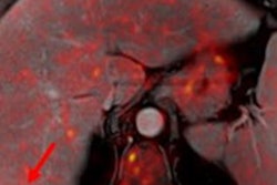
The scanner offers high-end MRI exams using conventional MRI, pre- and postcontrast, diffusion, perfusion, spectroscopy, and susceptibility when applicable, as well as PET data that uses the different tracers available based on a patient's disease, Dr. Yasser Abd El-Azim, co-founder and head of MRI at the private center, said.
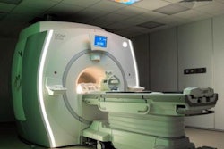
 The PET/MR scanner at the Misr Radiology Center. Copyright of the MSC.
"Hybrid imaging will be a great asset in the era we are entering into, where artificial intelligence specially deep learning and its vast applications, coinciding with molecular and genetic evaluations for various diseases are becoming a crucial part of diagnosis, treatment and follow-up, especially for cancer patients," he commented in an Q&A interview.
PET/MRI is well suited for the present and future examinations with its advanced ability of providing one stop shop exam offering the highest quality and the most accurate data, especially for cancer, epilepsy and dementia patients, he said.
"Hybrid imaging will continue to grow, and this would need more physicians to be able to interpret these advanced combined exams. Radiologists and nuclear medicine physicians need to work together more and we need to encourage and promote the development of hybrid subspecialty where radiologists and nuclear medicine physicians can join, to be trained to interpret, at least the main parts of both sides of the examination, he noted in the ESHIMT interview.