A group of German researchers has received the Image of the Year award from the Society of Nuclear Medicine and Molecular Imaging (SNMMI) for a series of PET images that show that older adults who maintained peak cognitive skills exhibited greater resistance to buildup of tau and amyloid proteins.
This group of cognitive overachievers was dubbed "super-agers" by the researchers, led by Merle Hoenig, PhD, from Research Center Juelich and University Hospital Cologne. They showed less tau and amyloid deposition on PET images than their counterparts in their 80s, who either had normal cognition or only had mild cognitive impairment.
"The phenomenon of super-aging suggests that cognitively high-functioning individuals have extraordinary mechanisms that resist brain aging processes and neurodegeneration," Hoenig said in a statement. "We know that tau pathology is more closely associated with cognitive decline than amyloid pathology. Thus, the resistance, in particular against tau pathology, likely allows these individuals to perform cognitively above average even at advanced age."
The researchers took subjects from the Alzheimer's Disease Neuroimaging Initiative (ADNI) to create three groups of 25 super-agers (median age, 85.2 ± 3.5 years), 25 normal-agers (median age, 84.5 ± 3.5 years), and 25 patients with mild cognitive impairment (median age, 84.7 ± 3.9 years). In addition, 18 cognitively normal, amyloid-negative controls (median age, 63.6 ± 2.7 years) were included as a reference group. Data included individual cognitive performances through ADNI composite memory scores over a four-year period prior to the PET scans.
PET imaging was performed with both F-18 AV-1451 (Flortaucipir, Avid Radiopharmaceuticals) and F-18 AV-45 (Florbetapir, Avid Radiopharmaceuticals) to compare the degree of tau and amyloid deposition among the four groups.
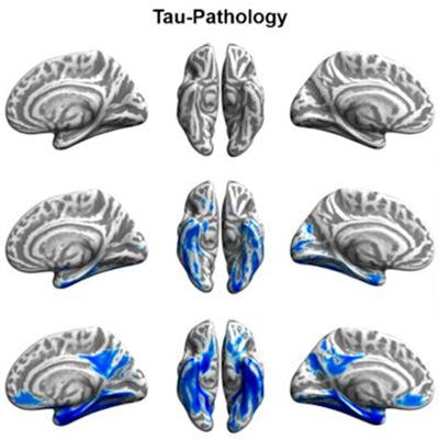
The PET images revealed no significant differences between the super-agers and the younger control group in terms of in vivo tau and amyloid burden. The normal-agers group exhibited tau burden in inferior temporal and precuneal areas but no significant differences in amyloid burden when compared with the controls. Patients with mild cognitive impairment showed both high amyloid and high tau pathology burden.
 Tau (blue) and amyloid (orange) distribution patterns for super-agers, normal-agers, and MCI patients, who were compared to a group of younger, healthy, cognitively normal, amyloid-negative individuals. Brain projections are depicted at an uncorrected significance (p < 0.0001). Color bars represent the respective t-statistic. Images courtesy of SNMMI and Hoenig et al.
The cognitive prowess of the super-agers "appears to be associated with the resistance to tau and amyloid pathology, which likely permits maintenance of cognitive performance despite advanced age," Hoenig and colleagues concluded. "In turn, differences between normal aging and mild cognitive impairment appear to be driven by the level of amyloid burden. These results motivate further research to determine responsible resistance factors, which may also inspire the development of novel treatment concepts."
As for the potential clinical ramifications, the researchers suggested that it will be challenging to develop therapeutics to tackle the multitude of factors involved in the aging process.
"However, if we understand which individuals are resistant to dementia, this will help us identify potential pathways that promote successful aging [and] protecting against not only Alzheimer's disease but also other aging-associated diseases, such as vascular disease and other forms of dementia," Hoenig added.
SNMMI's 2020 Image of the Year, which was chosen from more than 2,000 abstracts submitted to the meeting, "provides us with insight into how we can use these PET imaging biomarkers to understand behaviors and therapies that may allow more of us to age better and retain more of our cognitive abilities as we get older," said Dr. Umar Mahmood, PhD, chair of SNMMI's scientific program committee.
In 2019, the Image of the Year award was won by another group from Germany, specifically University Hospital Heidelberg, for their image of a gallium-68-based radiotracer that can detect 28 different types of cancer.