COVID-19_FDG_PET_CT_JNM_Albano_Image_G_400.png?auto=format%2Ccompress&q=70&w=400)
Nuclear medicine practitioners should be prepared for incidental findings of COVID-19 when patients undergo FDG-PET/CT scans for routine oncologic indications, according to a study published online on 1 April in the Journal of Nuclear Medicine.
Italian researchers reported that several patients who entered their facility asymptomatic for the novel coronavirus later showed signs of pneumonia on CT and FDG avidity on PET, which made them suspicious for COVID-19. By acting upon the incidental detection of COVID-19 pneumonia in its early stages, subsequent preemptive steps could help prevent the spread of the virus.
"Depending on local and contemporary regulations, an asymptomatic patient may stay at home with adequate clinical monitoring; in case of clinical doubts, more aggressive measures may be appropriate with the risk to postpone oncological treatments potentially affecting survival," wrote the authors, led by Dr. Domenico Albano from University of Brescia and Spedali Civili Brescia.
Italy has been especially devastated by the COVID-19 outbreak, with more than 114,000 cases and approximately 14,000 deaths as of 2 April. Initial symptoms include fever, cough, and trouble breathing, but other people who contract the virus remain asymptomatic. This seemingly healthy group who are unaware that they carry the virus could unknowingly spread it.
"While nuclear medicine procedures are unlikely to play a role in the primary diagnosis of COVID-19, there may be incidental detection in asymptomatic but infected cases undergoing scans for other indications, which may have relevant implications for further management," the authors added.
Between 16 March and 24 March, the nuclear medicine department in Brescia performed 65 FDG PET/CT scans (Discovery ST or 690 scanner, GE Healthcare) for routine oncological indications. All the patients were asymptomatic for the COVID-19 virus, but they were carefully screened before their admittance to the nuclear medicine unit, given the outbreak of SARS-CoV-2 infections in northern Italy. No reason was found to suspect COVID-19 at that time.
Following completion of the scans, six patients (9%) showed signs of pneumonia on CT and FDG avidity in all areas of pneumonia, which made these patients suspicious for COVID-19. The six patients included four women, between 55 and 65 years old, and two men, ages 65 and 77, respectively. When physicians followed up with reverse transcription polymerase chain reaction (RT-PCR) tests, five patients (83%) were confirmed to have COVID-19.
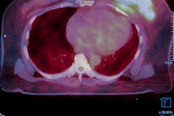
 Images include high-resolution thoracic CT with no sign of pneumonia (A, B). PET/CT performed one week later, showing bilateral pneumonia with ground-glass opacities at CT (C, D), PET (E, F), and PET/CT fused images (G, H) with SUVmax of five. Maximum intensity projection (I) shows axillary nodal lesion as FDG positive. Images courtesy of Journal of Nuclear Medicine.
To ensure the six cases of incidental pneumonia in March 2020 were not an aberration, the researchers compared their caseload from the same eight days of March 2019 and found only two cases (2.5%) of incidental pneumonia among 80 PET/CT studies.
"It should be noted, however, that our report is observatory in nature, and that a precise definition of the accuracy of PET findings of COVID would require prospective validation in larger cohorts," Albano and colleagues wrote.
Still, the researchers emphasized that it is "important to consider potential COVID-19 related findings during reading and to report such findings to the patient and his referring physicians immediately for appropriate action."