In a study covering more than 1,000 confirmed cases of the novel coronavirus disease (COVID-19), chest CT proved to be more accurate than the standard method of DNA testing for early diagnosis of the disease in high-risk individuals. The findings were published online on 26 February in Radiology.
Nucleic acid testing with reverse transcription polymerase chain reaction (RT-PCR) is considered the standard technique for diagnosing COVID-19. However, the long processing time and shaky outcomes of initial RT-PCR tests have made evident the need for a more efficient alternative for early diagnosis of the disease.
A recent examination of 51 cases demonstrated that CT had better sensitivity at detecting COVID-19 than RT-PCR testing among high-risk individuals upon initial presentation of symptoms, highlighting CT's utility in early diagnosis. The Chinese National Health Commission also sanctioned diagnosis through clinical symptoms and chest CT as an official method to confirm COVID-19 in the highly affected province of Hubei.
Expanding upon this prior work, researchers from China and the Netherlands examined data from 1,014 patients in Wuhan city, the presumed source of the outbreak. The patients were suspected of having COVID-19 and underwent a chest CT exam and had RT-PCR testing of throat swab samples between 6 January and 6 February.
Two radiologists with access to the patients' epidemiological history and clinical symptoms but blinded to the RT-PCR results classified the chest CT scans as positive or negative for COVID-19 based on the presence of key abnormal CT features. All of the patients were eventually confirmed to have COVID-19 with RT-PCR testing. The average age of the cohort was 51 years, and 56% were female.
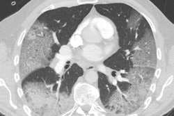
Axial chest CT images of a 62-year-old man with COVID-19 showing disease progression over time, from bilateral ground-glass opacities on 27 January (A) to multifocal organizing consolidation on 13 February (D). He had multiple negative results from RT-PCR tests, including those obtained on 3 and 11 February. Image courtesy of the RSNA.Overall, the researchers found that chest CT was better at diagnosing COVID-19 than RT-PCR testing upon initial examination of high-risk individuals. The initial RT-PCR testing period included multiple repeat tests up to three days after presentation.
Initial RT-PCR testing detected COVID-19 in only 59% of the cases. In comparison, 88% of the patients showed chest CT findings indicative of COVID-19. Among those with negative initial RT-PCR results, 93% showed CT features consistent with COVID-19 diagnosis prior to eventual confirmation on RT-PCR four or more days after the initial test.
Chest CT achieved a sensitivity of 97%, a specificity of 25%, and an accuracy of 68% for diagnosis of COVID-19 upon initial presentation of high-risk individuals, where eventual confirmation on RT-PCR served as the reference standard. The positive predictive value for chest CT was 65% and the negative predictive value was 83%.
"Our data and analysis suggest that chest CT should be considered for COVID-19 screening, comprehensive evaluation, and follow-up, especially in epidemic areas with high pretest probability for disease," wrote the authors, led by Dr. Tao Ai, PhD, and Dr. Zhenlu Yang, PhD, from Huazhong University of Science and Technology.
Relying on a combination of exposure history, clinical symptoms, and CT features rather than RT-PCR tests alone would aid in more rapid detection of the disease and may allow for better control of viral spread, they concluded.


![Examples of ultrasound findings and techniques. (A) Images in a 39-year-old male patient with a mass in the left thigh. The mass is heterogeneous on the B-mode US image (compared with the patient in D) and showed increased microvascularity (superb microvascular imaging [SMI]) and shear-wave elastography (SWE) values. Undifferentiated pleomorphic sarcoma was diagnosed at biopsy (with pleomorphic rhabdomyosarcoma in surgical specimen). (B) Images in an 18-year-old male patient with a mass in the left leg. The mass is hypoechoic on the B-mode image, with no other findings suggestive of malignancy. The lesion is in contact with the cortex of the tibia, which is slightly irregular. CT revealed a doubtful anteromedial tibial erosion. The microvascular study demonstrated high vascularization, suggestive of malignancy. Periosteal Ewing sarcoma was diagnosed with both histologic and immunohistochemical confirmation. (C) Images in a 69-year-old female patient with a lump growing on the outside of the left leg. Multiple SWE examinations were performed (please note the high values obtained in the measurements, whereas the color map highlights the stiffness relative to adjacent tissues). SMI showed areas of increased vascularization to target for sampling. Undifferentiated spindle cell sarcoma was diagnosed at biopsy, with residual leiomyosarcoma in the surgical specimen after neoadjuvant therapy. (D) Images in a 56-year-old female patient with a mass in the right thigh. The mass is heterogeneous at both B-mode ultrasound (similar to patient A) and MRI (coronal T2-weighted spectral attenuated inversion recovery [SPAIR]; T1-weighted pre-contrast and postcontrast imaging), which even shows uptake after the administration of paramagnetic contrast material, which is traditionally suggestive of malignancy. Low values at SMI and elastography are suggestive of benignity. Spindle cell lipoma was diagnosed at biopsy, with atypical spindle cell lipomatous tumor in the surgical specimen.](https://img.auntminnieeurope.com/mindful/smg/workspaces/default/uploads/2026/08/images-radiol250278fig2.APCFLSvX6p.jpg?auto=format%2Ccompress&fit=crop&h=100&q=70&w=100)


![Examples of ultrasound findings and techniques. (A) Images in a 39-year-old male patient with a mass in the left thigh. The mass is heterogeneous on the B-mode US image (compared with the patient in D) and showed increased microvascularity (superb microvascular imaging [SMI]) and shear-wave elastography (SWE) values. Undifferentiated pleomorphic sarcoma was diagnosed at biopsy (with pleomorphic rhabdomyosarcoma in surgical specimen). (B) Images in an 18-year-old male patient with a mass in the left leg. The mass is hypoechoic on the B-mode image, with no other findings suggestive of malignancy. The lesion is in contact with the cortex of the tibia, which is slightly irregular. CT revealed a doubtful anteromedial tibial erosion. The microvascular study demonstrated high vascularization, suggestive of malignancy. Periosteal Ewing sarcoma was diagnosed with both histologic and immunohistochemical confirmation. (C) Images in a 69-year-old female patient with a lump growing on the outside of the left leg. Multiple SWE examinations were performed (please note the high values obtained in the measurements, whereas the color map highlights the stiffness relative to adjacent tissues). SMI showed areas of increased vascularization to target for sampling. Undifferentiated spindle cell sarcoma was diagnosed at biopsy, with residual leiomyosarcoma in the surgical specimen after neoadjuvant therapy. (D) Images in a 56-year-old female patient with a mass in the right thigh. The mass is heterogeneous at both B-mode ultrasound (similar to patient A) and MRI (coronal T2-weighted spectral attenuated inversion recovery [SPAIR]; T1-weighted pre-contrast and postcontrast imaging), which even shows uptake after the administration of paramagnetic contrast material, which is traditionally suggestive of malignancy. Low values at SMI and elastography are suggestive of benignity. Spindle cell lipoma was diagnosed at biopsy, with atypical spindle cell lipomatous tumor in the surgical specimen.](https://img.auntminnieeurope.com/mindful/smg/workspaces/default/uploads/2026/08/images-radiol250278fig2.APCFLSvX6p.jpg?auto=format%2Ccompress&fit=crop&h=112&q=70&w=112)