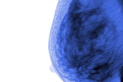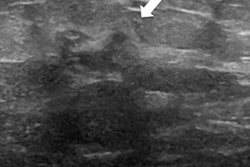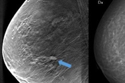
Italian researchers found that the combination of digital breast tomosynthesis (DBT) and ultrasound automated breast volume scanning (ABVS) measured up well against breast MRI for staging breast cancer. The findings were published in the October issue of the European Journal of Radiology.
The results suggest there's another tool in the arsenal for staging the disease, especially when MRI is unavailable or unfeasible, wrote a team led by Dr. Rossano Girometti of the University of Udine.
"MRI is the most sensitive tool to achieve local staging of breast cancer, with a significant impact on the type and extent of surgical treatment," the researchers wrote. "However, the use of MRI in the preoperative setting is still under debate, given limited availability, costs, and controversies on the low specificity reporting in previous [research]."
"In this light, local staging of breast cancer is frequently achieved using ultrasound coupled with digital mammography and/or DBT," they noted.
Because DBT has shown promise as a standalone adjunct to ultrasound, the group sought to investigate whether the combination of DBT and ABVS would prove an even more effective option (Eur J Radiol, October 2018, Vol. 107, pp. 188-195).
"ABVS has been introduced as a novel ultrasound-based modality to provide standardized scans of the breasts and a high-resolution volumetric set of images readable at any time. ... [However,] the role for ABVS in staging breast cancer has been poorly assessed, with a few works showing that this technique outperforms ultrasound in assessing breast cancer size ... or guiding breast conservative surgery in patients with ductal carcinoma in situ (DCIS)," the authors wrote.
The study included 73 breast cancer patients who underwent preoperative DBT, ABVS, and MRI between July 2015 and July 2016. Girometti and colleagues used histology and one-year follow-up data as their reference standard to calculate accuracy for the two strategies, comparing the DBT-ABVS combination with breast MRI.
There were 160 lesions in the patient cohort, 108 of which were cancer. The combination of DBT plus ABVS was comparable to MRI for all cancers, the researchers found, although it showed lower sensitivity and negative predictive value.
| Performance of DBT + ABVS vs. MRI for breast cancer | ||
| Measure | MRI | DBT + ABVS |
| Sensitivity | 96.3% | 91.7% |
| Specificity | 88.5% | 86.5% |
| Positive predictive value | 94.5% | 93.4% |
| Negative predictive value | 92% | 83.3% |
| Accuracy | 93.8% | 90% |
Although breast MRI outperformed the DBT-ABVS combination, the latter showed acceptable accuracy in staging breast cancer and could be used if MRI is unavailable, the group noted.
"To our knowledge, our study is the first to report that the direct combination of DBT and ABVS achieves comparable sensitivity (91.7%) and specificity (86.5%) [to breast MRI] in the same clinical setting," Girometti and colleagues concluded.


![Examples of ultrasound findings and techniques. (A) Images in a 39-year-old male patient with a mass in the left thigh. The mass is heterogeneous on the B-mode US image (compared with the patient in D) and showed increased microvascularity (superb microvascular imaging [SMI]) and shear-wave elastography (SWE) values. Undifferentiated pleomorphic sarcoma was diagnosed at biopsy (with pleomorphic rhabdomyosarcoma in surgical specimen). (B) Images in an 18-year-old male patient with a mass in the left leg. The mass is hypoechoic on the B-mode image, with no other findings suggestive of malignancy. The lesion is in contact with the cortex of the tibia, which is slightly irregular. CT revealed a doubtful anteromedial tibial erosion. The microvascular study demonstrated high vascularization, suggestive of malignancy. Periosteal Ewing sarcoma was diagnosed with both histologic and immunohistochemical confirmation. (C) Images in a 69-year-old female patient with a lump growing on the outside of the left leg. Multiple SWE examinations were performed (please note the high values obtained in the measurements, whereas the color map highlights the stiffness relative to adjacent tissues). SMI showed areas of increased vascularization to target for sampling. Undifferentiated spindle cell sarcoma was diagnosed at biopsy, with residual leiomyosarcoma in the surgical specimen after neoadjuvant therapy. (D) Images in a 56-year-old female patient with a mass in the right thigh. The mass is heterogeneous at both B-mode ultrasound (similar to patient A) and MRI (coronal T2-weighted spectral attenuated inversion recovery [SPAIR]; T1-weighted pre-contrast and postcontrast imaging), which even shows uptake after the administration of paramagnetic contrast material, which is traditionally suggestive of malignancy. Low values at SMI and elastography are suggestive of benignity. Spindle cell lipoma was diagnosed at biopsy, with atypical spindle cell lipomatous tumor in the surgical specimen.](https://img.auntminnieeurope.com/mindful/smg/workspaces/default/uploads/2026/08/images-radiol250278fig2.APCFLSvX6p.jpg?auto=format%2Ccompress&fit=crop&h=100&q=70&w=100)


![Examples of ultrasound findings and techniques. (A) Images in a 39-year-old male patient with a mass in the left thigh. The mass is heterogeneous on the B-mode US image (compared with the patient in D) and showed increased microvascularity (superb microvascular imaging [SMI]) and shear-wave elastography (SWE) values. Undifferentiated pleomorphic sarcoma was diagnosed at biopsy (with pleomorphic rhabdomyosarcoma in surgical specimen). (B) Images in an 18-year-old male patient with a mass in the left leg. The mass is hypoechoic on the B-mode image, with no other findings suggestive of malignancy. The lesion is in contact with the cortex of the tibia, which is slightly irregular. CT revealed a doubtful anteromedial tibial erosion. The microvascular study demonstrated high vascularization, suggestive of malignancy. Periosteal Ewing sarcoma was diagnosed with both histologic and immunohistochemical confirmation. (C) Images in a 69-year-old female patient with a lump growing on the outside of the left leg. Multiple SWE examinations were performed (please note the high values obtained in the measurements, whereas the color map highlights the stiffness relative to adjacent tissues). SMI showed areas of increased vascularization to target for sampling. Undifferentiated spindle cell sarcoma was diagnosed at biopsy, with residual leiomyosarcoma in the surgical specimen after neoadjuvant therapy. (D) Images in a 56-year-old female patient with a mass in the right thigh. The mass is heterogeneous at both B-mode ultrasound (similar to patient A) and MRI (coronal T2-weighted spectral attenuated inversion recovery [SPAIR]; T1-weighted pre-contrast and postcontrast imaging), which even shows uptake after the administration of paramagnetic contrast material, which is traditionally suggestive of malignancy. Low values at SMI and elastography are suggestive of benignity. Spindle cell lipoma was diagnosed at biopsy, with atypical spindle cell lipomatous tumor in the surgical specimen.](https://img.auntminnieeurope.com/mindful/smg/workspaces/default/uploads/2026/08/images-radiol250278fig2.APCFLSvX6p.jpg?auto=format%2Ccompress&fit=crop&h=112&q=70&w=112)