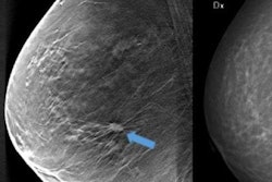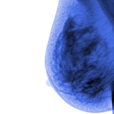
The addition of computer-aided detection (CAD) to digital breast tomosynthesis (DBT) interpretation can reduce reading time by 14% -- for both beginner and experienced readers, according to a study published online on 13 December in European Radiology.
The findings suggest that CAD is a useful tool when utilized with DBT, which can increase reading time due to the fact that it produces a 3D image, wrote a team led by Dr. Eun Young Chae of the University of Ulsan College of Medicine in Seoul, South Korea.
"Interpretation of DBT studies may be challenging for radiologists partially because it involves the analysis of a series of thin slices through the breast, which requires longer reading time than that associated with FFDM [full-field digital mammography]," the authors wrote. "To address the challenges of reviewing DBT studies, computer-aided detection software may help radiologists detect cancers and improve efficiency in the DBT interpretation workflow."
Because little research has been conducted exploring the use of CAD with DBT, Chae's group evaluated a DBT-CAD system they developed for the study to assess DBT interpretation and reading time between novice and experienced readers.
They included 100 DBT cases, 70 of which were malignant and 30 of which were benign. Most patients had heterogeneously dense or extremely dense tissue (82%). Four radiologists read the exams -- two experienced and two novice -- and each reader had two sessions separated by a two-month interval, one with CAD and one without. The researchers compared each reader's performance using area under the receiver operating characteristic curve (AUC) values and tracked reading time for each case.
They found no statistically significant difference in AUC for DBT exam interpretations for BI-RADS scale categories with CAD (0.778) and without CAD (0.776). However, reading times for both experienced and beginner readers decreased by 14% when CAD was used, from 72 seconds to 62 seconds. This reading time reduction was seen in both novice and experienced readers, the group noted.
| Average DBT exam interpretation time by reader, without and with CAD | |||
| Reader | Reading time without CAD in seconds | Reading time with CAD in seconds | p-value |
| Novice | 70.8 | 62 | 0.007 |
| Experienced | 73.3 | 62 | < 0.001 |
| All | 72 | 62 | < 0.001 |
Adding CAD to DBT exam interpretation improves reading time without negatively affecting the diagnostic performance of the radiologist.
"Although the impact of adding DBT to FFDM on the diagnostic performance may vary according to the experience level of the readers, the effect of CAD on DBT interpretation could be consistent regardless of their experience," Chae and colleagues concluded. "In this study, the novice readers performed as well as the experienced readers in all aspects with and without CAD."


![Examples of ultrasound findings and techniques. (A) Images in a 39-year-old male patient with a mass in the left thigh. The mass is heterogeneous on the B-mode US image (compared with the patient in D) and showed increased microvascularity (superb microvascular imaging [SMI]) and shear-wave elastography (SWE) values. Undifferentiated pleomorphic sarcoma was diagnosed at biopsy (with pleomorphic rhabdomyosarcoma in surgical specimen). (B) Images in an 18-year-old male patient with a mass in the left leg. The mass is hypoechoic on the B-mode image, with no other findings suggestive of malignancy. The lesion is in contact with the cortex of the tibia, which is slightly irregular. CT revealed a doubtful anteromedial tibial erosion. The microvascular study demonstrated high vascularization, suggestive of malignancy. Periosteal Ewing sarcoma was diagnosed with both histologic and immunohistochemical confirmation. (C) Images in a 69-year-old female patient with a lump growing on the outside of the left leg. Multiple SWE examinations were performed (please note the high values obtained in the measurements, whereas the color map highlights the stiffness relative to adjacent tissues). SMI showed areas of increased vascularization to target for sampling. Undifferentiated spindle cell sarcoma was diagnosed at biopsy, with residual leiomyosarcoma in the surgical specimen after neoadjuvant therapy. (D) Images in a 56-year-old female patient with a mass in the right thigh. The mass is heterogeneous at both B-mode ultrasound (similar to patient A) and MRI (coronal T2-weighted spectral attenuated inversion recovery [SPAIR]; T1-weighted pre-contrast and postcontrast imaging), which even shows uptake after the administration of paramagnetic contrast material, which is traditionally suggestive of malignancy. Low values at SMI and elastography are suggestive of benignity. Spindle cell lipoma was diagnosed at biopsy, with atypical spindle cell lipomatous tumor in the surgical specimen.](https://img.auntminnieeurope.com/mindful/smg/workspaces/default/uploads/2026/08/images-radiol250278fig2.APCFLSvX6p.jpg?auto=format%2Ccompress&fit=crop&h=100&q=70&w=100)


![Examples of ultrasound findings and techniques. (A) Images in a 39-year-old male patient with a mass in the left thigh. The mass is heterogeneous on the B-mode US image (compared with the patient in D) and showed increased microvascularity (superb microvascular imaging [SMI]) and shear-wave elastography (SWE) values. Undifferentiated pleomorphic sarcoma was diagnosed at biopsy (with pleomorphic rhabdomyosarcoma in surgical specimen). (B) Images in an 18-year-old male patient with a mass in the left leg. The mass is hypoechoic on the B-mode image, with no other findings suggestive of malignancy. The lesion is in contact with the cortex of the tibia, which is slightly irregular. CT revealed a doubtful anteromedial tibial erosion. The microvascular study demonstrated high vascularization, suggestive of malignancy. Periosteal Ewing sarcoma was diagnosed with both histologic and immunohistochemical confirmation. (C) Images in a 69-year-old female patient with a lump growing on the outside of the left leg. Multiple SWE examinations were performed (please note the high values obtained in the measurements, whereas the color map highlights the stiffness relative to adjacent tissues). SMI showed areas of increased vascularization to target for sampling. Undifferentiated spindle cell sarcoma was diagnosed at biopsy, with residual leiomyosarcoma in the surgical specimen after neoadjuvant therapy. (D) Images in a 56-year-old female patient with a mass in the right thigh. The mass is heterogeneous at both B-mode ultrasound (similar to patient A) and MRI (coronal T2-weighted spectral attenuated inversion recovery [SPAIR]; T1-weighted pre-contrast and postcontrast imaging), which even shows uptake after the administration of paramagnetic contrast material, which is traditionally suggestive of malignancy. Low values at SMI and elastography are suggestive of benignity. Spindle cell lipoma was diagnosed at biopsy, with atypical spindle cell lipomatous tumor in the surgical specimen.](https://img.auntminnieeurope.com/mindful/smg/workspaces/default/uploads/2026/08/images-radiol250278fig2.APCFLSvX6p.jpg?auto=format%2Ccompress&fit=crop&h=112&q=70&w=112)