Developing diagnostic reference levels (DRLs) for common CT procedures in an orthopedic clinical setting, along with an emphasis on education and training of CT radiographers, are key factors in the optimization process, Irish researchers have asserted.
During the onsite training in the CT department at the National Orthopaedic Hospital Cappagh (NOHC) in Dublin, radiographers learn about the importance of minimizing the scan area to the required anatomical regions only and to avoid unnecessary repeat scanning. Each patient request is carefully reviewed before scanning commences to ensure the correct protocols and scan areas are selected, according to consultant radiologist Prof. Stephen Eustace and his colleagues at the NOHC.
Considerable effort has also gone into the development of local DRLs, and the researchers hope these DRLs will assist other hospitals carrying out similar procedures in their optimization of patient exposures. “Previous to this study, there were no benchmark DRLs for these CT procedures for comparison. The DRLs will be used in the NOHC to further optimize local CT imaging protocols and assist staff in keeping patient doses as low as reasonably achievable,” they noted in a EuroSafe 2025 e-poster.
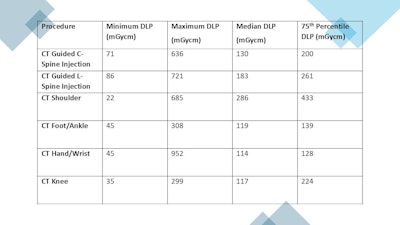 CT diagnostic reference levels (DRLs) developed at the National Orthopaedic Hospital Cappagh. DLP = dose length product.All figures courtesy of M. Smyth, B. Byrne, L. Gavagan, and S. Eustace, presented at EuroSafe 2025.
The NOHC installed its first CT scanner (Definition Edge from Siemens Healthineers) in October 2021. Before this installation, patients requiring CT scans were transferred to other acute hospitals for imaging, which resulted in delayed diagnostics and placed additional pressures on the acute hospital system.
The new machine facilitated musculoskeletal (MSK) tumour biopsies, ablations, and minimally invasive spinal procedures. It also made possible diagnostic tumour staging, coronary and pulmonary CT angiography, and prosthesis modular design planning aided by metal artifact reduction software. By early 2025, over 4,000 patients had been scanned on this unit, which is Ireland’s only CT scanner dedicated to orthopedic imaging, the authors stated.
Medical exposure to ionizing radiation is one of the largest contributors to artificial ionizing radiation exposure for the Irish population, accounting for 10.4% of total exposure. CT accounts for 62% of the radiation dose from all medical exposure in Ireland, but only 11% of the procedures are completed, according to the Ionising Radiation National Dose Report.
Against this background, the authors’ three main goals were to establish DRLs for common CT procedures in an orthopedic clinical setting, to establish DRLs for two CT MSK interventional procedures, and to aid optimization of CT procedures in a dedicated orthopedic clinical setting.
DRLs are dose levels set to aid optimization of diagnostic and interventional procedures, and they provide a standard for comparison to help ensure the radiation protection of patients undergoing different types of medical radiological procedures, they explained. Under EU and Irish legislation, each hospital/facility is required to review its DRLs on a regular basis. In Ireland, national DRLs exist for the most commonly performed CT examinations.
During the annual review of the DRLs for the CT scanner at the NOHC, Eustace and his colleagues found that there were no national or international DRLs for some of the CT procedures and examinations carried out in the NOHC. A literature review found a lack of published DRLs for common CT examinations in an orthopedic clinical setting. The NOHC is working toward EuroSafe Imaging Star recognition, so the team decided to establish its own DRLs based on data collected over the previous three years.
The group reviewed workload in the CT scanner and decided to select a mixture of CT-guided interventional procedures and commonly requested anatomy for inclusion in the study. Cervical facet joint and lumbar spine joint injections are commonly carried out on the CT scanner by a number of different radiologists.
“Radiologists usually remain outside of the scan room during these procedures in order to keep their radiation exposure to a minimum,” they wrote. “Four body parts commonly scanned were chosen for inclusion in the study: shoulder, knees, foot/ankle, and hand/wrist CT scans.”
For each examination, approximately 100 patients who were scanned using the typical protocol were included in the analysis. The DRLs were presented based on the dose length product (DLP) for the CT scan, the median and 75th percentile being quoted for each procedure.
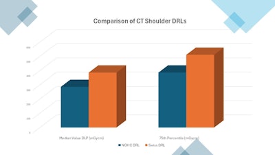 Comparison of shoulder diagnostic reference levels (DRLs).
As indicated in the bar graph, the CT DRL for shoulder examinations established by the Irish team was approximately 70% of that established in a Swiss study (R. Treier et al. Patient doses in CT examinations in Switzerland: implementation of national diagnostic reference levels. Radiation Protection Dosimetry, Volume 142, Issue 2-4, December 2010, pp 244-254).
Software provided by the scanner manufacturer, such as the CARE Dose4D CT automatic exposure control system and CARE kV (a fully automated feature integrated into the workflow at the CT scanner that optimizes tube voltage), are routinely used at the NOHC to achieve significant dose reductions based on the individual patient’s size and shape, the authors pointed out.
Due to the nature of orthopedic imaging and the presence of many metallic implants, iMAR (iterative Metal Artifact Reduction) is used. This software helps to reduce artifacts caused by implants, artificial joints, and pacemakers, they added.
You can access the full EuroSafe 2025 e-poster on the ESR’s EPOS website.


