
A dental brace wire was found in a woman's bowel -- 10 years after she had last worn dental braces, according to a new case report published online in BMJ Case Reports (7 August 2017).
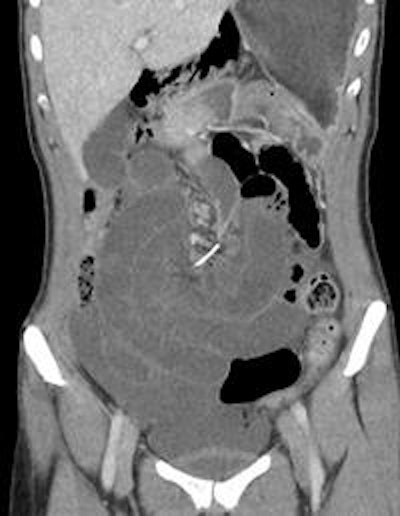
Coronal CT image of orthodontic wire at the root of the small-bowel volvulus. Image courtesy of BMJ Case Reports.
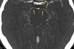
After an initial visit to the emergency department (ED) where she had been thought to have biliary colic, a previously healthy 30-year-old woman returned to the ED two days later complaining of worsening central abdominal pain, according to the group from Sir Charles Gairdner Hospital in Nedlands, Western Australia. A CT scan of the abdomen showed a metallic, wire-shaped foreign body at the mesenteric root of a small-bowel volvulus. The wire had pierced several parts of the small bowel, causing the volvulus.
The patient hadn't worn braces for 10 years and didn't recall ingesting the wire or noticing that her braces wire was missing. The authors noted that foreign body ingestion should be considered as a cause of abdominal pain in patients with no other medical or surgical history.
"Routine plain film of the abdomen is a useful initial investigation and should not be overlooked," the authors wrote.
The full case report can be found here.


![Overview of the study design. (A) The fully automated deep learning framework was developed to estimate body composition (BC) (defined as subcutaneous adipose tissue [SAT] in liters; visceral adipose tissue [VAT] in liters; skeletal muscle [SM] in liters; SM fat fraction [SMFF] as a percentage; and intramuscular adipose tissue [IMAT] in deciliters) from MRI. The fully automated framework comprised one model (model 1) to quantify different BC measures (SAT, VAT, SM, SMFF, and IMAT) as three-dimensional (3D) measures from whole-body MRI scans. The second model (model 2) was trained to identify standardized anatomic landmarks along the craniocaudal body axis (z coordinate field), which allowed for subdividing the whole-body measures into different subregions typically examined on clinical routine MRI scans (chest, abdomen, and pelvis). (B) BC was quantified from whole-body MRI in over 66,000 individuals from two large population-based cohort studies, the UK Biobank (UKB) (36,317 individuals) and the German National Cohort (NAKO) (30,291 individuals). Bar graphs show age distribution by sex and cohort. BMI = body mass index. (C) After the performance assessment of the fully automated framework, the change in BC measures, distributions, and profiles across age decades were investigated. Age-, sex-, and height-adjusted body composition reference curves were calculated and made publicly available in a web-based z-score calculator (https://circ-ml.github.io).](https://img.auntminnieeurope.com/mindful/smg/workspaces/default/uploads/2026/05/body-comp.XgAjTfPj1W.jpg?auto=format%2Ccompress&fit=crop&h=112&q=70&w=112)


