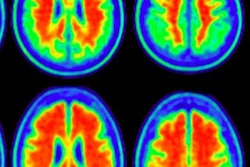
While PET can accurately detect or exclude Alzheimer's disease by imaging beta-amyloid plaques in the brain, the modality is underutilized in clinical care, according to research presented this week at the Alzheimer's Association International Conference (AAIC) in London.
PET brain scans have proved more effective for evaluating Alzheimer's disease in a larger proportion of patients than standard structural and metabolic imaging and cerebrospinal fluid (CSF) assessment. In addition, using PET to detect the presence of beta-amyloid plaques can change diagnoses in as many as two-thirds of cases.
"A negative brain PET scan indicating sparse to no amyloid plaques rules out Alzheimer's disease as the cause of dementia symptoms. This makes it a valuable tool to clarify an uncertain or difficult diagnosis," said James Hendrix, PhD, director of global science initiatives for the Alzheimer's Association, in a statement. "Misdiagnosis is costly to health systems and expensive and distressing to persons with dementia and their families."
In one ongoing study, researchers at Oslo University Hospital, led by Dr. Nenad Bogdanovic, PhD, found that PET amyloid imaging was a key contributor to diagnosing or excluding Alzheimer's disease in all 50 study participants. In contrast, CSF testing for amyloid allowed for diagnosis or exclusion in 44 (88%) of the 50 individuals using a higher detection cutoff and in only 21 individuals (42%) using traditional cutoffs.
Meanwhile, in a study in Sweden, PET imaging prompted a change in diagnosis in a strong majority of participants. Antoine Leuzy, a doctoral student at the Karolinska Institute in Stockholm, and colleagues evaluated the use of PET in 61 individuals diagnosed with mild cognitive impairment, Alzheimer's disease, other types of dementia, or severe cognitive impairment.
Leuzy et al found that PET imaging led to a change in diagnosis in 68% of these participants. Meanwhile, agreement between CSF testing and PET imaging was only 53% to 57%, depending on the approach used to read the PET scan.
Other PET-related research presented at AAIC 2017 included a study on access to PET imaging. U.S. researchers surveyed 510 participants and caregivers and found that over 80% were dissatisfied with the availability of PET scans, supported additional research with the modality, and would undergo PET imaging if the scans were recommended by their physician.
Another study, an analysis of Medicare claims data, found that approximately 60% of dementia cases are missed in clinical practice, particularly cases of early dementia.


![Examples of ultrasound findings and techniques. (A) Images in a 39-year-old male patient with a mass in the left thigh. The mass is heterogeneous on the B-mode US image (compared with the patient in D) and showed increased microvascularity (superb microvascular imaging [SMI]) and shear-wave elastography (SWE) values. Undifferentiated pleomorphic sarcoma was diagnosed at biopsy (with pleomorphic rhabdomyosarcoma in surgical specimen). (B) Images in an 18-year-old male patient with a mass in the left leg. The mass is hypoechoic on the B-mode image, with no other findings suggestive of malignancy. The lesion is in contact with the cortex of the tibia, which is slightly irregular. CT revealed a doubtful anteromedial tibial erosion. The microvascular study demonstrated high vascularization, suggestive of malignancy. Periosteal Ewing sarcoma was diagnosed with both histologic and immunohistochemical confirmation. (C) Images in a 69-year-old female patient with a lump growing on the outside of the left leg. Multiple SWE examinations were performed (please note the high values obtained in the measurements, whereas the color map highlights the stiffness relative to adjacent tissues). SMI showed areas of increased vascularization to target for sampling. Undifferentiated spindle cell sarcoma was diagnosed at biopsy, with residual leiomyosarcoma in the surgical specimen after neoadjuvant therapy. (D) Images in a 56-year-old female patient with a mass in the right thigh. The mass is heterogeneous at both B-mode ultrasound (similar to patient A) and MRI (coronal T2-weighted spectral attenuated inversion recovery [SPAIR]; T1-weighted pre-contrast and postcontrast imaging), which even shows uptake after the administration of paramagnetic contrast material, which is traditionally suggestive of malignancy. Low values at SMI and elastography are suggestive of benignity. Spindle cell lipoma was diagnosed at biopsy, with atypical spindle cell lipomatous tumor in the surgical specimen.](https://img.auntminnieeurope.com/mindful/smg/workspaces/default/uploads/2026/08/images-radiol250278fig2.APCFLSvX6p.jpg?auto=format%2Ccompress&fit=crop&h=100&q=70&w=100)


![Examples of ultrasound findings and techniques. (A) Images in a 39-year-old male patient with a mass in the left thigh. The mass is heterogeneous on the B-mode US image (compared with the patient in D) and showed increased microvascularity (superb microvascular imaging [SMI]) and shear-wave elastography (SWE) values. Undifferentiated pleomorphic sarcoma was diagnosed at biopsy (with pleomorphic rhabdomyosarcoma in surgical specimen). (B) Images in an 18-year-old male patient with a mass in the left leg. The mass is hypoechoic on the B-mode image, with no other findings suggestive of malignancy. The lesion is in contact with the cortex of the tibia, which is slightly irregular. CT revealed a doubtful anteromedial tibial erosion. The microvascular study demonstrated high vascularization, suggestive of malignancy. Periosteal Ewing sarcoma was diagnosed with both histologic and immunohistochemical confirmation. (C) Images in a 69-year-old female patient with a lump growing on the outside of the left leg. Multiple SWE examinations were performed (please note the high values obtained in the measurements, whereas the color map highlights the stiffness relative to adjacent tissues). SMI showed areas of increased vascularization to target for sampling. Undifferentiated spindle cell sarcoma was diagnosed at biopsy, with residual leiomyosarcoma in the surgical specimen after neoadjuvant therapy. (D) Images in a 56-year-old female patient with a mass in the right thigh. The mass is heterogeneous at both B-mode ultrasound (similar to patient A) and MRI (coronal T2-weighted spectral attenuated inversion recovery [SPAIR]; T1-weighted pre-contrast and postcontrast imaging), which even shows uptake after the administration of paramagnetic contrast material, which is traditionally suggestive of malignancy. Low values at SMI and elastography are suggestive of benignity. Spindle cell lipoma was diagnosed at biopsy, with atypical spindle cell lipomatous tumor in the surgical specimen.](https://img.auntminnieeurope.com/mindful/smg/workspaces/default/uploads/2026/08/images-radiol250278fig2.APCFLSvX6p.jpg?auto=format%2Ccompress&fit=crop&h=112&q=70&w=112)