Dear Advanced Visualization Insider,
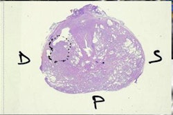
Computer-aided detection (CAD) is now being used in a number of anatomic regions, but unfortunately the prostate hasn't been one of them. Until now, as researchers are turning in their first studies of prostate CAD based on artificial intelligence (AI) and deep learning.
Looking at about 100 patients with suspected prostate cancer, researchers from the University of Rome applied their AI-based CAD system to MR images of potential tumor regions identified by expert readers, hoping to improve the yield of core biopsies to detect malignancy. Find out what happened by clicking here.
Not to be outdone, a team from China presenting at the ECR 2017 evaluated deep-learning techniques for reading prostate MRI. Click here to find out how well they performed and what the complications were.
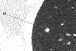
In another CAD story, researchers from Japan analyzed lung CT images and found that using CAD in combination with advanced iterative reconstruction techniques allowed even small pulmonary nodules to be seen at sharply reduced doses. They found limits to this practice, however, which you can learn about here.
Another group from Italy put lung CAD against human readers to compare detection rates for subsolid pulmonary nodules. Care to guess who won?
In head CT, a team from the Netherlands developed a color mapping technique to help pinpoint areas of ischemia in acute stroke imaging. As predicted, use of the technique sped up interpretation in a setting where time is brain.
Looking at the bigger picture for a moment, what are radiologists to do about increasing workloads and declining reimbursement? Why, artificial intelligence of course, which contributing writer Stephen Holloway calls the "great enabler" for an era in which so much in radiology seems to be getting more complicated. In fact, he shows AI can address a wide range of problems and complications from back-office bottlenecks to interpretation dilemmas that slow workflow. He presents the evidence here.
We invite you to scroll through the links below to find the rest of the news in advanced visualization, 3D visualization and printing, and computer-aided detection -- brought to you fresh daily in your Advanced Visualization Community.


![Examples of ultrasound findings and techniques. (A) Images in a 39-year-old male patient with a mass in the left thigh. The mass is heterogeneous on the B-mode US image (compared with the patient in D) and showed increased microvascularity (superb microvascular imaging [SMI]) and shear-wave elastography (SWE) values. Undifferentiated pleomorphic sarcoma was diagnosed at biopsy (with pleomorphic rhabdomyosarcoma in surgical specimen). (B) Images in an 18-year-old male patient with a mass in the left leg. The mass is hypoechoic on the B-mode image, with no other findings suggestive of malignancy. The lesion is in contact with the cortex of the tibia, which is slightly irregular. CT revealed a doubtful anteromedial tibial erosion. The microvascular study demonstrated high vascularization, suggestive of malignancy. Periosteal Ewing sarcoma was diagnosed with both histologic and immunohistochemical confirmation. (C) Images in a 69-year-old female patient with a lump growing on the outside of the left leg. Multiple SWE examinations were performed (please note the high values obtained in the measurements, whereas the color map highlights the stiffness relative to adjacent tissues). SMI showed areas of increased vascularization to target for sampling. Undifferentiated spindle cell sarcoma was diagnosed at biopsy, with residual leiomyosarcoma in the surgical specimen after neoadjuvant therapy. (D) Images in a 56-year-old female patient with a mass in the right thigh. The mass is heterogeneous at both B-mode ultrasound (similar to patient A) and MRI (coronal T2-weighted spectral attenuated inversion recovery [SPAIR]; T1-weighted pre-contrast and postcontrast imaging), which even shows uptake after the administration of paramagnetic contrast material, which is traditionally suggestive of malignancy. Low values at SMI and elastography are suggestive of benignity. Spindle cell lipoma was diagnosed at biopsy, with atypical spindle cell lipomatous tumor in the surgical specimen.](https://img.auntminnieeurope.com/mindful/smg/workspaces/default/uploads/2026/08/images-radiol250278fig2.APCFLSvX6p.jpg?auto=format%2Ccompress&fit=crop&h=100&q=70&w=100)


![Examples of ultrasound findings and techniques. (A) Images in a 39-year-old male patient with a mass in the left thigh. The mass is heterogeneous on the B-mode US image (compared with the patient in D) and showed increased microvascularity (superb microvascular imaging [SMI]) and shear-wave elastography (SWE) values. Undifferentiated pleomorphic sarcoma was diagnosed at biopsy (with pleomorphic rhabdomyosarcoma in surgical specimen). (B) Images in an 18-year-old male patient with a mass in the left leg. The mass is hypoechoic on the B-mode image, with no other findings suggestive of malignancy. The lesion is in contact with the cortex of the tibia, which is slightly irregular. CT revealed a doubtful anteromedial tibial erosion. The microvascular study demonstrated high vascularization, suggestive of malignancy. Periosteal Ewing sarcoma was diagnosed with both histologic and immunohistochemical confirmation. (C) Images in a 69-year-old female patient with a lump growing on the outside of the left leg. Multiple SWE examinations were performed (please note the high values obtained in the measurements, whereas the color map highlights the stiffness relative to adjacent tissues). SMI showed areas of increased vascularization to target for sampling. Undifferentiated spindle cell sarcoma was diagnosed at biopsy, with residual leiomyosarcoma in the surgical specimen after neoadjuvant therapy. (D) Images in a 56-year-old female patient with a mass in the right thigh. The mass is heterogeneous at both B-mode ultrasound (similar to patient A) and MRI (coronal T2-weighted spectral attenuated inversion recovery [SPAIR]; T1-weighted pre-contrast and postcontrast imaging), which even shows uptake after the administration of paramagnetic contrast material, which is traditionally suggestive of malignancy. Low values at SMI and elastography are suggestive of benignity. Spindle cell lipoma was diagnosed at biopsy, with atypical spindle cell lipomatous tumor in the surgical specimen.](https://img.auntminnieeurope.com/mindful/smg/workspaces/default/uploads/2026/08/images-radiol250278fig2.APCFLSvX6p.jpg?auto=format%2Ccompress&fit=crop&h=112&q=70&w=112)