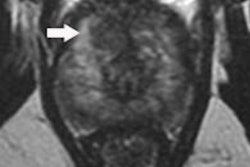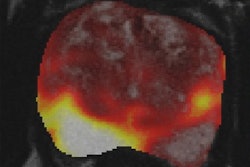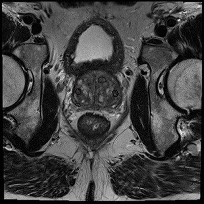
By first undergoing a multiparametric MRI scan, more than 25% of men with suspected prostate cancer could avoid a needle biopsy guided by transrectal ultrasound (TRUS), according to a study published online January 19 in Lancet.
In addition, the ability of multiparametric MRI to more precisely locate and gauge the size of tumors improves the detection of aggressive cancers when combined with a follow-up biopsy, concluded researchers from the University College London (UCL) Hospitals NHS Foundation Trust (Lancet, January 19, 2017).
 Diagnostic accuracy for detection of clinically significant cancer (primary definition) between MP-MRI and TPM-biopsy. MP-MRI = multiparametric MRI. TPM-biopsy = template prostate mapping biopsy. Pie charts represent actual MP-MRI scores 1-5. Sensitivity 93% (95% CI 88-96), positive predictive value 51% (46-56), specificity 41% (36-46), negative predictive value 89% (83-94). All figures courtesy of the Lancet.
The findings are from the Prostate MR Imaging Study (PROMIS), under the guidance of the Medical Research Council (MRC) Clinical Trials Unit at the UCL.
TRUS biopsies often follow high readings of prostate-specific antigen (PSA) in blood tests. The procedure involves the insertion of an ultrasound probe into the patient's rectum to guide the biopsy needle into the prostate to take 10 to 12 small tissue samples.
Each year some 100,000 men in the U.K. have a TRUS biopsy. Approximately 66% of the results are negative for cancer or indicate no life-threatening cancer. The invasive procedure, however, has side effects, including pain, bleeding, erectile dysfunction, and infection. The results may also be inaccurate because the TRUS biopsy takes samples from various areas of the prostate in a random manner.
In PROMIS, the researchers analyzed 576 men with suspected prostate cancer at 11 NHS hospitals who received a multiparametric MRI scan followed by two types of biopsy. The first procedure was a template prostate mapping biopsy, which was used as a control to compare the accuracy of the multiparametric MRI and standard biopsy. A TRUS biopsy was then performed.
 Diagnostic accuracy for detection of clinically significant cancer (primary definition) between TRUS-biopsy and TPM-biopsy. TRUS-biopsy = transrectal ultrasound-guided prostate biopsy. TPM-biopsy = template prostate mapping biopsy. Sensitivity 48% (95% CI 42-55), positive predictive value 90% (83-94), specificity 96% (94-98), negative predictive value 74% (69-78).
Multiparametric MRI scans detected 93% of clinically important cancers, while TRUS biopsies identified only 48% of clinically important cancers. In addition, 89% of subjects who had a negative multiparametric MRI scan had either no cancer or a harmless cancer.
Dr. Hashim Ahmed, one of the lead investigators from the UCL, said the results clearly show that having an MRI scan followed by a TRUS biopsy can dramatically improve the diagnosis of prostate cancer. The approach could also help around one-quarter of men with negative MRI scans to avoid having an immediate biopsy and its potential side effects, he added.
The researchers also noted that more information is needed regarding the overall cost-effectiveness of this strategy, and preparatory work should be conducted to ensure there are enough MRI scanners and appropriately trained urologists and radiologists available to further implement the approach.
Editor's note: Image on the home page courtesy of Geert Litjens, Radboud University Medical Center in Nijmegen, the Netherlands.