Researchers at Trinity College in Dublin have devised an experimental 3D bone scanning technique that may improve the diagnosis of bone injuries without the use of ionizing radiation, according to a new study published in Chemical Communications.
The technique begins with researchers attaching luminescent compounds to tiny gold structures to form nanoagents that are attracted to calcium-rich surfaces. These structures appear when bones form even the tiniest cracks, the authors noted in a statement.
The biologically safe nanoagents target and highlight the cracks formed in damaged bones, allowing researchers to produce a complete 3D image of the damaged regions (Chemical Communications, September 2016, Vol. 18-52:72, pp. 10,858-10,861).
Daily bone damage
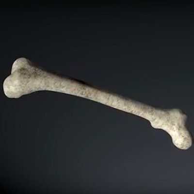
Everyday activity stresses the bones and causes microcracks to develop, explained Dr. Clive Lee, professor of anatomy in the Royal College of Surgeons in Ireland (RCSI) and the Royal Hibernian Academy and visiting professor of biomechanics in Trinity College Dublin. Bones are normally repaired by a modeling process, but when the microcracks develop too fast they can grow faster than the remodeling process and ultimately weaken bones, he added. The process occurs in athletes and in the elderly, who develop especially extensive fractures when osteoporosis is present.
The current imaging standard, x-ray, can see the bone and major fractures but provides little information about bone structure and quality. By using the nanoagent to label microcracks and detecting them with MRI, the team hopes to study bone quality and quantity and deliver appropriate therapy, especially regarding the diagnoses of weak bones before they break.
The high resolution of the technique is a major advantage, along with the lack of radiation, the researchers said.
The project comes from many years of collaboration between Trinity chemists and engineering experts from the RCSI.
The technique results in a highly detailed 3D map of bone damage showing microcracks using noninvasive luminescence imaging, the authors wrote. With the nanoagent, bone damage can be demonstrated in a manner that wasn't possible previously, and the work represents a major step in the development of targeted contrast agents for use in clinical applications.
Science Foundation Ireland funded the project, which also benefited from collaboration with scientists at RCSI, led by Lee.