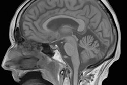
Researchers from the Medical University of Vienna in Austria have used neuroimaging techniques to uncover important biological differences in the brains of men and women, as well as those who identify as transsexual.
After performing MRI on transsexual subjects receiving hormonal treatment and on control subjects, the researchers found sex hormones had a significant impact on brain connections and functions.
"For clinical research, this means we may get better insights into which hormonal dysfunctions may be linked to anxiety disorders and why women suffer from such disorders two to three times more often than men," principal investigator Dr. Rupert Lanzenberger, PhD, said in a statement. "The more knowledge we have about gender differences, the better medical science will be able to target therapeutic measures precisely, and the better the advances in so-called personalized medicine will be."
In an attempt to explore biological factors in mental illnesses, the researchers studied transsexual subjects with MRI to observe what happens in the brain when opposite-sex hormones are administered over a prolonged period in order to achieve opposite-sex hormonal blood levels. They also imaged control subjects.
Over the five-year study, the team found the hormones had an effect on language processing, risk-taking behavior, spatial cognition, impulsiveness, and structural brain connections. They also determined that transgender individuals had a brain structure with levels falling in the mid-range between the two sexes.
For example, an increase in blood testosterone levels resulted in a decrease in the volume of two brain regions that are of central importance for language processing and it also changed their connections, according to the group.
"This suggests the impact of testosterone on language processing occurs via the influence it has on the structure of gray and white matter in the corresponding brain region," Lanzenberger said. "We assume that some of the difference in white matter we found may emerge very early on, perhaps in the womb or before puberty. That would make it a type of biological information, a marker for gender identity."
The researchers believe the knowledge that brain connections and functions can change as a result of hormone administration even in adulthood may be important, such as when brain neuroplasticity is reduced in certain situations, for instance as presumed in depression. Using PET imaging, the group also found that testosterone significantly increased serotonin transporter density.
In ongoing investigations, the Vienna researchers said they have recorded a large volume of data on pain sensitivity, the sense of smell, behavior changes, and quality of life in order to correlate them with the brain scans. The goal is ultimately gain a better understanding of gender differences, according to the group.
The research was supported by the Austria Science Fund FWF.


![Examples of ultrasound findings and techniques. (A) Images in a 39-year-old male patient with a mass in the left thigh. The mass is heterogeneous on the B-mode US image (compared with the patient in D) and showed increased microvascularity (superb microvascular imaging [SMI]) and shear-wave elastography (SWE) values. Undifferentiated pleomorphic sarcoma was diagnosed at biopsy (with pleomorphic rhabdomyosarcoma in surgical specimen). (B) Images in an 18-year-old male patient with a mass in the left leg. The mass is hypoechoic on the B-mode image, with no other findings suggestive of malignancy. The lesion is in contact with the cortex of the tibia, which is slightly irregular. CT revealed a doubtful anteromedial tibial erosion. The microvascular study demonstrated high vascularization, suggestive of malignancy. Periosteal Ewing sarcoma was diagnosed with both histologic and immunohistochemical confirmation. (C) Images in a 69-year-old female patient with a lump growing on the outside of the left leg. Multiple SWE examinations were performed (please note the high values obtained in the measurements, whereas the color map highlights the stiffness relative to adjacent tissues). SMI showed areas of increased vascularization to target for sampling. Undifferentiated spindle cell sarcoma was diagnosed at biopsy, with residual leiomyosarcoma in the surgical specimen after neoadjuvant therapy. (D) Images in a 56-year-old female patient with a mass in the right thigh. The mass is heterogeneous at both B-mode ultrasound (similar to patient A) and MRI (coronal T2-weighted spectral attenuated inversion recovery [SPAIR]; T1-weighted pre-contrast and postcontrast imaging), which even shows uptake after the administration of paramagnetic contrast material, which is traditionally suggestive of malignancy. Low values at SMI and elastography are suggestive of benignity. Spindle cell lipoma was diagnosed at biopsy, with atypical spindle cell lipomatous tumor in the surgical specimen.](https://img.auntminnieeurope.com/mindful/smg/workspaces/default/uploads/2026/08/images-radiol250278fig2.APCFLSvX6p.jpg?auto=format%2Ccompress&fit=crop&h=100&q=70&w=100)


![Examples of ultrasound findings and techniques. (A) Images in a 39-year-old male patient with a mass in the left thigh. The mass is heterogeneous on the B-mode US image (compared with the patient in D) and showed increased microvascularity (superb microvascular imaging [SMI]) and shear-wave elastography (SWE) values. Undifferentiated pleomorphic sarcoma was diagnosed at biopsy (with pleomorphic rhabdomyosarcoma in surgical specimen). (B) Images in an 18-year-old male patient with a mass in the left leg. The mass is hypoechoic on the B-mode image, with no other findings suggestive of malignancy. The lesion is in contact with the cortex of the tibia, which is slightly irregular. CT revealed a doubtful anteromedial tibial erosion. The microvascular study demonstrated high vascularization, suggestive of malignancy. Periosteal Ewing sarcoma was diagnosed with both histologic and immunohistochemical confirmation. (C) Images in a 69-year-old female patient with a lump growing on the outside of the left leg. Multiple SWE examinations were performed (please note the high values obtained in the measurements, whereas the color map highlights the stiffness relative to adjacent tissues). SMI showed areas of increased vascularization to target for sampling. Undifferentiated spindle cell sarcoma was diagnosed at biopsy, with residual leiomyosarcoma in the surgical specimen after neoadjuvant therapy. (D) Images in a 56-year-old female patient with a mass in the right thigh. The mass is heterogeneous at both B-mode ultrasound (similar to patient A) and MRI (coronal T2-weighted spectral attenuated inversion recovery [SPAIR]; T1-weighted pre-contrast and postcontrast imaging), which even shows uptake after the administration of paramagnetic contrast material, which is traditionally suggestive of malignancy. Low values at SMI and elastography are suggestive of benignity. Spindle cell lipoma was diagnosed at biopsy, with atypical spindle cell lipomatous tumor in the surgical specimen.](https://img.auntminnieeurope.com/mindful/smg/workspaces/default/uploads/2026/08/images-radiol250278fig2.APCFLSvX6p.jpg?auto=format%2Ccompress&fit=crop&h=112&q=70&w=112)