Dear Advanced Visualization Insider,
Traumatic injuries and illnesses -- including cancer -- mean there will always be high demand for mandible reconstruction. But until now, clinicians have had to get by with rough visual estimates of missing mandible sections to plan reconstructive surgery and select appropriate bone graft donors.
A team of researchers from Aachen University in Germany may have found a better way. They developed a technique that enables the reconstruction of mandibles accurately in about three minutes by using a new topology-based approach. The protocol is accurate enough to complete even large continuity defects. How did they do it? Click here.
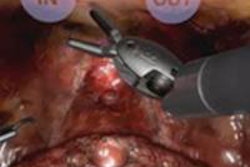
Meanwhile, investigators from Israel have dedicated themselves to developing virtual training modules for difficult interventional procedures. By creating a development platform that can be modified to demonstrate a broad range of procedures, the group has taken steps to ensure that surgeons and interventionalists will have plenty of practice before they try their skills on human patients. Get the nuts and bolts of this exciting research project by clicking here.
You've surely seen some of those impressive functional MRI (fMRI) studies in which researchers demonstrate what parts of the brain allow us to lift a pencil, jump for joy, or play Mozart on the piano. But parts of the brain that make us worry may be activated when folks learn that fMRI software can be surprisingly inaccurate, according to new results from the U.K. The study team logged false brain activity in more than half the cases, thanks to faulty brain-mapping software. This means tens of thousands of fMRI studies may be off the mark. Get the rest of the story here.
Correct or not, however, the use of medical imaging analysis software to diagnose myriad diseases is a burgeoning industry, according to a recent market report.
Finally, machine-learning technology is being deployed to uncover the mysteries of Alzheimer's disease, according to new research from the Netherlands. The Amsterdam-based investigators used machine-learning techniques to analyze arterial spin-labeled MRI scans. The application was trained to recognize patterns to distinguish between patients with varying levels of cognitive impairment and to predict the stage of Alzheimer's disease cases that were not yet diagnosed.
For the rest of the news in your Advanced Visualization Community, we invite you to scroll through the links below.


![Examples of ultrasound findings and techniques. (A) Images in a 39-year-old male patient with a mass in the left thigh. The mass is heterogeneous on the B-mode US image (compared with the patient in D) and showed increased microvascularity (superb microvascular imaging [SMI]) and shear-wave elastography (SWE) values. Undifferentiated pleomorphic sarcoma was diagnosed at biopsy (with pleomorphic rhabdomyosarcoma in surgical specimen). (B) Images in an 18-year-old male patient with a mass in the left leg. The mass is hypoechoic on the B-mode image, with no other findings suggestive of malignancy. The lesion is in contact with the cortex of the tibia, which is slightly irregular. CT revealed a doubtful anteromedial tibial erosion. The microvascular study demonstrated high vascularization, suggestive of malignancy. Periosteal Ewing sarcoma was diagnosed with both histologic and immunohistochemical confirmation. (C) Images in a 69-year-old female patient with a lump growing on the outside of the left leg. Multiple SWE examinations were performed (please note the high values obtained in the measurements, whereas the color map highlights the stiffness relative to adjacent tissues). SMI showed areas of increased vascularization to target for sampling. Undifferentiated spindle cell sarcoma was diagnosed at biopsy, with residual leiomyosarcoma in the surgical specimen after neoadjuvant therapy. (D) Images in a 56-year-old female patient with a mass in the right thigh. The mass is heterogeneous at both B-mode ultrasound (similar to patient A) and MRI (coronal T2-weighted spectral attenuated inversion recovery [SPAIR]; T1-weighted pre-contrast and postcontrast imaging), which even shows uptake after the administration of paramagnetic contrast material, which is traditionally suggestive of malignancy. Low values at SMI and elastography are suggestive of benignity. Spindle cell lipoma was diagnosed at biopsy, with atypical spindle cell lipomatous tumor in the surgical specimen.](https://img.auntminnieeurope.com/mindful/smg/workspaces/default/uploads/2026/08/images-radiol250278fig2.APCFLSvX6p.jpg?auto=format%2Ccompress&fit=crop&h=100&q=70&w=100)


![Examples of ultrasound findings and techniques. (A) Images in a 39-year-old male patient with a mass in the left thigh. The mass is heterogeneous on the B-mode US image (compared with the patient in D) and showed increased microvascularity (superb microvascular imaging [SMI]) and shear-wave elastography (SWE) values. Undifferentiated pleomorphic sarcoma was diagnosed at biopsy (with pleomorphic rhabdomyosarcoma in surgical specimen). (B) Images in an 18-year-old male patient with a mass in the left leg. The mass is hypoechoic on the B-mode image, with no other findings suggestive of malignancy. The lesion is in contact with the cortex of the tibia, which is slightly irregular. CT revealed a doubtful anteromedial tibial erosion. The microvascular study demonstrated high vascularization, suggestive of malignancy. Periosteal Ewing sarcoma was diagnosed with both histologic and immunohistochemical confirmation. (C) Images in a 69-year-old female patient with a lump growing on the outside of the left leg. Multiple SWE examinations were performed (please note the high values obtained in the measurements, whereas the color map highlights the stiffness relative to adjacent tissues). SMI showed areas of increased vascularization to target for sampling. Undifferentiated spindle cell sarcoma was diagnosed at biopsy, with residual leiomyosarcoma in the surgical specimen after neoadjuvant therapy. (D) Images in a 56-year-old female patient with a mass in the right thigh. The mass is heterogeneous at both B-mode ultrasound (similar to patient A) and MRI (coronal T2-weighted spectral attenuated inversion recovery [SPAIR]; T1-weighted pre-contrast and postcontrast imaging), which even shows uptake after the administration of paramagnetic contrast material, which is traditionally suggestive of malignancy. Low values at SMI and elastography are suggestive of benignity. Spindle cell lipoma was diagnosed at biopsy, with atypical spindle cell lipomatous tumor in the surgical specimen.](https://img.auntminnieeurope.com/mindful/smg/workspaces/default/uploads/2026/08/images-radiol250278fig2.APCFLSvX6p.jpg?auto=format%2Ccompress&fit=crop&h=112&q=70&w=112)