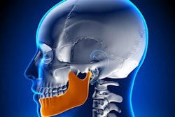
German researchers used a 3D advanced visualization algorithm to reconstruct mandibles with missing parts -- the first step in the process of mandible bone restoration in patients with severe illness or injury, they wrote in the International Journal of Computer-Assisted Radiology and Surgery.
Using volumetric data from 60 CT scans of the mandible as a guide, the investigators created a 3D visual mesh that follows the course of the jaw's anatomic structures to mimic the shape of the bone. Virtual reconstructions showed the results sufficiently accurate to allow surgical reconstruction planning, with average deviations of less than 2 mm for small missing parts and less than 5 mm for large hemilateral defects.
The technique "bears the potential of being used as the first essential step in a process chain of computer-aided manufacturing of bone scaffolds for mandibular reconstruction," explained Stefan Raith and colleagues from Aachen University Hospital in Germany. This is in contrast to how surgical planning is performed today, based mainly on visual judgment and the individual skills and experience of the surgeons (IJCARS, 8 July 2016).
Not for lack of trying
Although several methods of improving craniofacial reconstructive surgery have been proposed, no planning tool has yet found its way to routine use in the operating room, Raith and colleagues wrote. But the need is there.
Patients can lose portions of their mandibles as a result of ablation procedures or traumatic accidents. Lacking a computerized reconstruction scheme, surgeons find it difficult to estimate the shape of the missing portions and choose an appropriate graft donor to supply the missing part. In some cases, mirroring the healthy side of the bone is an option for providing the defined target geometry required for successful reconstruction, but this approach is not possible in every case.
That opens the door to computational-based methods for calculating target reconstruction shapes. Among previous attempts, Zachow et al introduced a method based on principal component analysis, dividing the mandibular surface into patches used to generate a statistical representation to replace severely malformed mandibles. Another group mapped mandibular geometry to a generic sphere to acquire a common parameterization for segmenting the shapes based on CT data. But this study is the first to use a topological representation that can be adjusted to the complex shape features of the mandible, the authors wrote.
The investigators introduced a new statistical shape representation of anatomy-related topology and demonstrated its suitability for planning reconstructive surgery in patients with large continuity defects of the mandible, furnishing individually optimized shapes, wrote Raith and colleagues.
"Our hypothesis is that it is possible with this technique to reconstruct the missing mandibular sections with an accuracy that meets the requirements of clinical practice," they wrote.
Mesh defined by vertices
The study relied on volumetric data from 63 CT datasets of mandibles from cadavers. The specimens, which lacked information on age, sex, or ethnicity, were scanned in a multidetector-row CT scanner (Brilliance iCT, Philips Healthcare). The volumetric CT data were segmented using Mimics 14.0 software (Materialise), and artifacts were removed as necessary, the authors wrote.
For topologically optimized surface representation, the native data segmentations were stored in a triangulated stereolithography (STL) file format. But because triangles cause topology mismatches and are not ideal for comparing similar surfaces, the researchers chose quadrilateral representation to convert the mandible surfaces to meshes, relying on single standardized surface topology. Quadrilaterals "allow a straightforward definition of a continuous edge flow that allows following of certain characteristic anatomical lines," the group wrote.
The semiautomated procedure requires manual user interactions for landmark placement. This was accomplished by defining a coarse mesh with 555 vertices or edge points. The design allows each vertex point to be projected toward the individual mesh -- a process that formed the subsequent modeling of all mandible shapes in the study, the group wrote. The graphic user interface was designed to allow geometric modification of the standardized surface by manipulating a small number of master points corresponding to salient anatomical points.
Further detailed manipulations to the mesh geometry were implemented by defining so-called dependent nodes, which are modified in tandem with the master nodes to enable fast refinement of coarse mandibular geometry, the authors wrote. The influence of both master and dependent nodes was applied using the same Laplacian surface editing approach.
The coarse mesh was refined, and surface projection operations automatically applied to the mesh until the mandible shape was represented by 8,649 vertices and 8,576 quadrilateral elements. The resulting mesh, implemented in Python programming language and executed in Blender open-source software in about three minutes, was smoothly distributed over the individual target geometry.
After converting segmented data to standardized mesh topology, the researchers performed an analysis to align the mandibles with optimal rotation and translation to a common mean, Raith and colleagues wrote.
Accurate for all sizes
Evaluation of the accuracy of the technique on six virtually defined continuity defects not included in the initial database showed virtual reconstructions accurate enough for surgical reconstruction planning, with average deviations toward the actual geometry of 1.82 ± 0.11 for small missing parts and 5 mm for large hemilateral defects, the group wrote.
"Goodness of fit with reference to the segmented raw data [was shown to be] excellent for the whole mandible surface," the team wrote.
The calculations passed visual inspections as well, revealing a natural appearance. Shapes were classified independently to be valid mandible shapes by three different experienced surgeons, the group noted.
Needs improvement
Still, the number of samples should be increased from 60 in future work, and demographic data on the samples could also improve the results, the authors noted.
"We expect that the accuracy of the method will be improved by such an increase in available data, as more special features and unusual mandible shapes may be present in the reconstruction database," they wrote. A cross-validation (or leave-one-out study) might be performed in future work to improve statistical analysis.
Also, even though the method is completely automated at the stage of reconstruction shape calculation, the algorithm architecture enables additional user input if desired, Raith and colleagues wrote. This would allow the surgeon to adjust the shapes of the reconstructed parts by applying adjusted factors to the calculated shape modes.
The quadrilateral mesh topology corresponded to special anatomical features of the mandible and followed its characteristic anatomical curves, the authors wrote. A further advantage of mesh representation is it allows the application of defined surgical bony incisions for tumor surgery modeling. Other approaches used for morphological studies on the mandible use generic geometries that do not offer this feature.
An advantage of the shape representation is its lack of special requirements concerning the topology of the original segmented surface data, "such as that it is a closed surface homeomorphic to a solid sphere," the team wrote. This allows teeth to be excluded and the alveolar ridge to be left open, improving virtual planning of dental implants.
Finally, they said, the method is not limited to the mandible, and may be applied in the future to a wide variety of anatomical structures.


