The University of Cambridge and Addenbrooke's Hospital in the U.K. said they have completed what they believe to be Europe's first metabolic imaging study using a new, hyperpolarized, MRI-contrast technique.
Developed after many years of research in collaboration with GE Healthcare, the hyperpolarized, MRI-contrast method will enable imaging of molecular changes in patients, offering potential applications in cancer detection and treatment monitoring, according to the group. The method involves labeling pyruvate -- a breakdown product of glucose that's found in all cells -- with carbon-13, according to the group. After the carbon atoms are hyperpolarized, they become at least 10,000 times more sensitive to detection in an MRI scanner, according to the team.
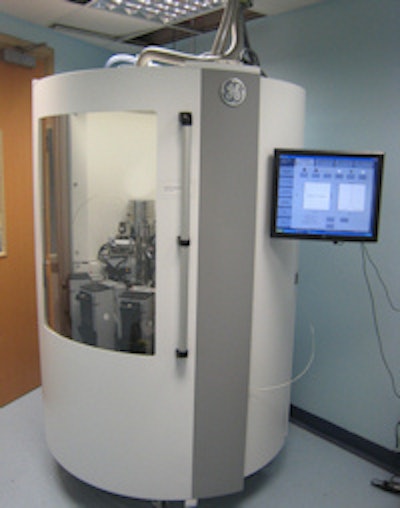
 The system used by the Cambridge team.
Once the pyruvate is injected into the patient, the signals can be tracked as the molecule moves around the body and enters cells, according to the group.
"MRI can then monitor how quickly the cells break it down and the rate at which [tumor] cells do this can tell the clinician whether or not a drug has been effective at killing them," the group said in a statement.
The Cambridge team said they recently imaged their first patient, demonstrating uptake of pyruvate in organs within the chest and abdomen. Previously, the imaging technique had only been used on patients in North America.
Professor Kevin Brindle of the Cancer Research UK Cambridge Institute and Dr. Ferdia Gallagher jointly led the project, which also included co-investigators and collaborators at the University of Cambridge and at Addenbrooke's Hospital. The facility and clinical trials are being funded and supported by the Wellcome Trust, Cancer Research UK, Addenbrooke's Charitable Trust, the Cambridge Cancer Centre, Cambridge University Hospitals National Health Service (NHS) Foundation Trust, GE, and the University of Cambridge.