Dear Molecular Imaging Insider,
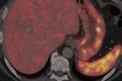
A new study has added to the growing evidence that PET/CT imaging with the radiopharmaceutical gallium-68 (Ga-68) DOTATATE should be the modality of choice for patients with neuroendocrine tumors.
German researchers found that Ga-68 DOTATATE-PET/contrast CT achieved greater sensitivity and specificity in the diagnostic accuracy and detection of bone metastases and lymph node metastases compared with contrast CT alone. It also changed the clinical management in more than 25% of patients in the study cohort. Read more by clicking here.
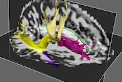
Meanwhile, U.K. researchers have used neuroimaging to confirm that meditation can create the same physiological changes in the brain as medication for patients with clinical depression. More research needs to be done, but initial evidence suggests that, instead of medicating, people should try meditating.
Young pediatric patients may benefit from FDG-PET/MRI for staging and clinically evaluating oncologic disorders, given the hybrid modality's superior soft-tissue contrast and significantly lower radiation dose. That's the view of German researchers who evaluated pediatric cancer patients younger than 6 years old, potentially changing the treatment plan for almost half of the sample.
PET/MRI can also provide high diagnostic performance for restaging gynecological cancer patients compared with FDG-PET/CT, with only slightly longer scan time but with "markedly reduced" radiation exposure. The benefits from PET/MRI were achieved along with no significant differences in sensitivity, specificity, positive predictive value, negative predictive value, and diagnostic accuracy, according to this German study.
A pan-European group is looking into the future and predicts that imaging is on the "cusp of a revolution" in both early and established dementia. They anticipate that within 10 years new imaging techniques, software, and biomarkers will assist specialists in diagnosing patients early and aid the development of much-needed treatments. They added that FDG-PET currently holds the most promise, given the vast number of trials using the modality for this clinical application.
There is always news in molecular imaging. Be sure to stay in touch with the Molecular Imaging Community on a daily basis to be informed on the latest developments.


![Examples of ultrasound findings and techniques. (A) Images in a 39-year-old male patient with a mass in the left thigh. The mass is heterogeneous on the B-mode US image (compared with the patient in D) and showed increased microvascularity (superb microvascular imaging [SMI]) and shear-wave elastography (SWE) values. Undifferentiated pleomorphic sarcoma was diagnosed at biopsy (with pleomorphic rhabdomyosarcoma in surgical specimen). (B) Images in an 18-year-old male patient with a mass in the left leg. The mass is hypoechoic on the B-mode image, with no other findings suggestive of malignancy. The lesion is in contact with the cortex of the tibia, which is slightly irregular. CT revealed a doubtful anteromedial tibial erosion. The microvascular study demonstrated high vascularization, suggestive of malignancy. Periosteal Ewing sarcoma was diagnosed with both histologic and immunohistochemical confirmation. (C) Images in a 69-year-old female patient with a lump growing on the outside of the left leg. Multiple SWE examinations were performed (please note the high values obtained in the measurements, whereas the color map highlights the stiffness relative to adjacent tissues). SMI showed areas of increased vascularization to target for sampling. Undifferentiated spindle cell sarcoma was diagnosed at biopsy, with residual leiomyosarcoma in the surgical specimen after neoadjuvant therapy. (D) Images in a 56-year-old female patient with a mass in the right thigh. The mass is heterogeneous at both B-mode ultrasound (similar to patient A) and MRI (coronal T2-weighted spectral attenuated inversion recovery [SPAIR]; T1-weighted pre-contrast and postcontrast imaging), which even shows uptake after the administration of paramagnetic contrast material, which is traditionally suggestive of malignancy. Low values at SMI and elastography are suggestive of benignity. Spindle cell lipoma was diagnosed at biopsy, with atypical spindle cell lipomatous tumor in the surgical specimen.](https://img.auntminnieeurope.com/mindful/smg/workspaces/default/uploads/2026/08/images-radiol250278fig2.APCFLSvX6p.jpg?auto=format%2Ccompress&fit=crop&h=100&q=70&w=100)


![Examples of ultrasound findings and techniques. (A) Images in a 39-year-old male patient with a mass in the left thigh. The mass is heterogeneous on the B-mode US image (compared with the patient in D) and showed increased microvascularity (superb microvascular imaging [SMI]) and shear-wave elastography (SWE) values. Undifferentiated pleomorphic sarcoma was diagnosed at biopsy (with pleomorphic rhabdomyosarcoma in surgical specimen). (B) Images in an 18-year-old male patient with a mass in the left leg. The mass is hypoechoic on the B-mode image, with no other findings suggestive of malignancy. The lesion is in contact with the cortex of the tibia, which is slightly irregular. CT revealed a doubtful anteromedial tibial erosion. The microvascular study demonstrated high vascularization, suggestive of malignancy. Periosteal Ewing sarcoma was diagnosed with both histologic and immunohistochemical confirmation. (C) Images in a 69-year-old female patient with a lump growing on the outside of the left leg. Multiple SWE examinations were performed (please note the high values obtained in the measurements, whereas the color map highlights the stiffness relative to adjacent tissues). SMI showed areas of increased vascularization to target for sampling. Undifferentiated spindle cell sarcoma was diagnosed at biopsy, with residual leiomyosarcoma in the surgical specimen after neoadjuvant therapy. (D) Images in a 56-year-old female patient with a mass in the right thigh. The mass is heterogeneous at both B-mode ultrasound (similar to patient A) and MRI (coronal T2-weighted spectral attenuated inversion recovery [SPAIR]; T1-weighted pre-contrast and postcontrast imaging), which even shows uptake after the administration of paramagnetic contrast material, which is traditionally suggestive of malignancy. Low values at SMI and elastography are suggestive of benignity. Spindle cell lipoma was diagnosed at biopsy, with atypical spindle cell lipomatous tumor in the surgical specimen.](https://img.auntminnieeurope.com/mindful/smg/workspaces/default/uploads/2026/08/images-radiol250278fig2.APCFLSvX6p.jpg?auto=format%2Ccompress&fit=crop&h=112&q=70&w=112)