In a small study published in Acta Paediatrica, researchers in the U.K. used 4D ultrasound to detect changes in facial movements of fetuses that could reflect the effect of maternal smoking.
Researchers at Durham and Lancaster universities, led by Dr. Nadja Reissland of Durham, performed 80 4D ultrasound scans on 20 fetuses to assess subtle mouth and touch movements. The scans were acquired at four different intervals between 24 and 36 weeks of pregnancy (Acta Paediatr, March 12, 2015).
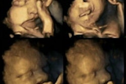
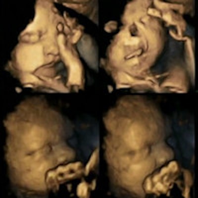
 4D ultrasound scans show movement differences in two fetuses at 32 weeks, above and below. The top strip of each image shows movements of a fetus whose mother smoked, while the bottom strip shows a fetus whose mother did not smoke. Images courtesy of Dr. Nadja Reissland, Durham University.
Four of the fetuses were carried by women who smoked an average of 14 cigarettes a day. The other 16 fetuses were carried by women who didn't smoke. All fetuses were healthy when born.
The researchers tracked mouth movements on the scans, and they found a statistically significant difference between the fetuses carried by the smoking women versus those of the nonsmokers (p < 0.02). The fetuses of the smoking women showed a higher rate of movements than the normal declining rate expected during pregnancy, and the differences between the smoking and nonsmoking groups widened as the pregnancies progressed.
The researchers speculated that the central nervous systems of the fetuses carried by the smoking women did not develop at the same rate and in the same way as in the fetuses of the women who didn't smoke. Other studies have reported delays in speech processing in infants exposed to smoke during pregnancy.