European researchers are investigating different phase-contrast x-ray tomography methods to find out which one might be the most useful for clinical applications.
Phase-contrast x-ray has the potential to produce better-quality images with more contrast than conventional x-ray imaging, which could help differentiate cancerous tissue from healthy tissue. Phase-contrast x-ray makes the x-ray beam interfere while it propagates from the target to a detector; this differs from conventional radiography, which instead relies on the attenuation of the amplitude of x-ray waves to produce images.
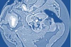
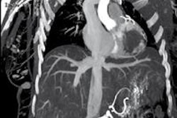
Researchers used three types of phase-contrast x-ray tomography methods to assess which one worked best: x-ray grating interferometry, propagation-based phase tomography with single-distance phase reconstruction, and holotomography.
They generated x-ray radiation using a synchrotron at the European Synchrotron Radiation Facility (ESRF) in France and tested each of the three techniques by examining cancerous tissue from a mouse and a rat's heart. Their results were reported this week in the Journal of Applied Physics.
For each specimen, holotomography and single-distance phase reconstruction produced spatial resolution that was twice as good as x-ray grating interferometry. But x-ray grating interferometry provided better contrast-to-noise ratios for anatomical features. It also excelled at density measurements and was more robust against low-frequency artifacts than holotomography.
The researchers concluded that all three methods were complementary, depending on the spatial and density resolutions required for the imaging task at hand, as well as the radiation dose requirements. The synchrotron experiments could provide useful benchmarks when translating phase-contrast tomography techniques to clinical practice, they concluded.