Using whole-body and micro CT, U.K. researchers have concluded that King Richard III suffered three injuries that potentially could have caused his death at the Battle of Bosworth Field in 1485, according to a study published online on 17 September in the Lancet.
The forensic analysis of the king's skeletal remains, led by the University of Leicester in the U.K., revealed two injuries to the skull and one injury to the pelvis that could have been deadly.
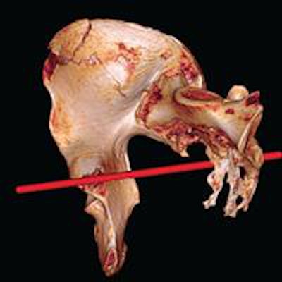
 Image of right hemi-pelvis and sacrum reconstructed from postmortem CT scan with Osirix software. Red line shows estimated direction of sharp-force trauma.
Whole-body CT scans and micro-CT imaging showed that Richard's now 500-year-old skeleton sustained 11 wounds. In total, there were nine skull injuries inflicted in battle, indicating that he may have removed or lost his helmet. There also were two injuries to the postcranial skeleton.
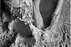
Researchers also analyzed tool marks on bone to identify the medieval weapons that may have been responsible for his injuries.
The research, led by Dr. Jo Appleby of the University of Leicester School of Archaeology and Ancient History, speculated that the postcranial injuries might have been inflicted after Richard's death. Had he been alive, he would have been wearing armor that would have prevented such wounds.
King Richard III's remains were found under a car park in Leicester by archaeologists from the University of Leicester. A multidisciplinary team from the university later connected the discovery to the king.