Radiologists have much to offer as volunteer physicians for Mercy Ships, a nonprofit organization that provides much-needed basic medical care to people in developing nations. In a recent article in the American Journal of Roentgenology, a U.S. radiologist who served on the Africa Mercy described his experiences.
In Africa alone, nearly half of the population has no access to a hospital or doctor, according to the global charity organization, which operates a fleet of hospital ships with an all-volunteer staff. The first radiologist aboard one of the ships was Dr. Robert Harris of Dartmouth-Hitchcock Medical Center, who served on the Africa Mercy for 10 days in 2012, when the ship was docked in Lomé, Togo (AJR, February 2013, Vol. 200:2, pp. W124-W129).
"I got involved with doing international work with ultrasound back in 2005, when I went to Serbia with one of our medical students, back to his hometown," Harris said. "We also have a Dartmouth group that goes to Nicaragua, and I took a portable ultrasound unit to a small hospital there. When I became aware of Mercy Ships I made an inquiry, and they said, 'Sure, we'd love to have you -- we've never had a radiologist on board.' "
 |
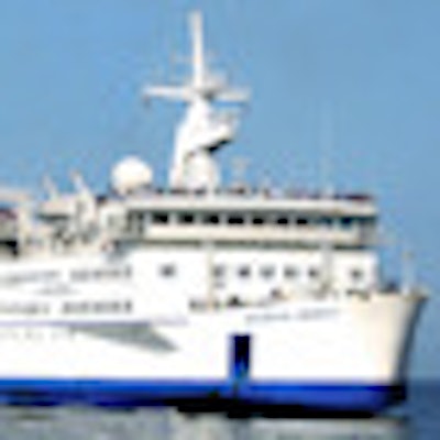
| The Africa Mercy. Image courtesy of Mercy Ships. |
Radiologists have much to offer, Harris said. Those with neurologic and head and neck experience who are also able to do some ultrasound, including neck and thyroid imaging, would be especially useful to surgeons performing maxillofacial procedures. And musculoskeletal radiologists or those with pediatric bone experience could support pediatric orthopedic surgeons.
Based in Lindale, TX, Mercy Ships has offered ship-board medical care around the world since 1978, according to the organization. In Africa, medical staff work with patients who present with conditions such as cleft lips or palates, cataracts, old trauma and orthopedic injuries, head and neck tumors, incontinence, and vesicovaginal fistulas. Although Mercy Ships staff does serve in emergencies, its main mission is to provide ongoing, specialized medical services to underserved populations.
The organization acquired the Africa Mercy, formerly a Danish rail ferry, in 1999. The ship has six operating theaters and a 78-bed ward; its annual medical capacity is approximately 7,000 surgical procedures onboard, according to Mercy Ships.
 Dr. Robert Harris.
The Africa Mercy's imaging resources include a plain radiography system; a computed radiography (CR) unit that allows for elongated films up to 82.5 cm for lower extremity or scoliosis views; a single-slice helical CT scanner; a compact ultrasound device with general curvilinear and high-resolution linear probes; a conebeam CT scanner used primarily in dental procedures with maxillofacial reconstruction surgery; and a panoramic x-ray machine. The ship also has a PACS.
The Africa Mercy's imaging capabilities are supplemented by volunteer radiologists in Canada, the U.S., the U.K., and Australia, who offer consultations to the ship's surgeons or internists, Harris wrote. However, most studies are not reviewed by a radiologist but are informally interpreted by the referring physician. To explain the radiographic or sonographic procedures to patients, Mercy Ships hires day workers from the native country where the ship is docked.
Radiologic technologists/radiographers in these types of settings often need additional resourcefulness and skills, Harris wrote. For example, technologists often receive CT requisitions that lack contrast or slice-thickness specifications, leaving them to determine scan protocols using their best judgment.
"As radiologists we have a lot of skills -- like setting CT protocols -- that we may not realize are valuable to people, because we do them every day," Harris said.
The average monthly radiology workload includes 160 radiography exams, 20 CT exams, 30 conebeam CT studies, 20 ultrasound exams, and 40 panoramic x-ray studies, Harris wrote.
"Most CT examinations are ordered for preoperative planning or to triage between surgery and nonsurgical treatment, but also to diagnose common problems among the crew or day workers, such as appendicitis or diverticulitis," he wrote. "Most of the plain radiographs, when I was onboard, were for day workers with positive purified protein derivative (ppd) placements, to rule out remote or active tuberculosis. All the studies I interpreted (approximately 10) were negative."
Costs to volunteer include airfare to the country being served and a nominal daily charge for room and board (about $30 to $40), according to Harris.
"Radiologists can enhance imaging services onboard the Africa Mercy ship, which does incredibly meaningful and even life-changing work for patients in the low-resource settings of West Africa," he said. "Contact Mercy Ships and choose a time to serve when there will be other doctors there who can use your expertise."
Information on volunteering is available at the organization's website, or by emailing the group at [email protected].