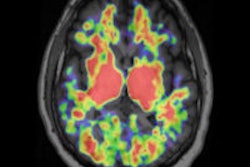
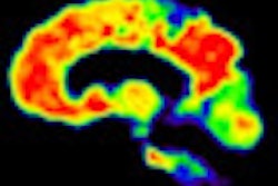
The National Health Service (NHS) in the U.K. now will cover PET beta-amyloid imaging scans to rule out Alzheimer's disease.
The decision marks the first time that PET beta-amyloid imaging will be covered by a public health system and adds validity to the imaging technique to diagnose dementia. The first-ever NHS patient to receive a PET beta-amyloid brain scan for Alzheimer's occurred at the Imperial Hospital Trust in London.
The NHS announcement came during December's G8 Dementia Summit as part of a plan to allocate as much as 66 million euros by 2015 for research into Alzheimer's. The Medical Research Council (MRC) also will invest 150 million euros for clinical research in the U.K., with 50 million euros to better understand how dementia affects the brain and improve early detection and treatments to delay progression of the disease.
In the U.S., the Centers for Medicare and Medicaid Services (CMS) in July 2013 reaffirmed its decision not to fully endorse PET beta-amyloid imaging for Medicare beneficiaries with dementia or neurodegenerative disease.
In its proposed decision memo, the agency ruled there is "insufficient" evidence to conclude that the use of PET beta-amyloid imaging improves health outcomes for patients with dementia or neurodegenerative disease. However, the agency agreed to cover one PET beta-amyloid scan per patient under certain circumstances.


![Overview of the study design. (A) The fully automated deep learning framework was developed to estimate body composition (BC) (defined as subcutaneous adipose tissue [SAT] in liters; visceral adipose tissue [VAT] in liters; skeletal muscle [SM] in liters; SM fat fraction [SMFF] as a percentage; and intramuscular adipose tissue [IMAT] in deciliters) from MRI. The fully automated framework comprised one model (model 1) to quantify different BC measures (SAT, VAT, SM, SMFF, and IMAT) as three-dimensional (3D) measures from whole-body MRI scans. The second model (model 2) was trained to identify standardized anatomic landmarks along the craniocaudal body axis (z coordinate field), which allowed for subdividing the whole-body measures into different subregions typically examined on clinical routine MRI scans (chest, abdomen, and pelvis). (B) BC was quantified from whole-body MRI in over 66,000 individuals from two large population-based cohort studies, the UK Biobank (UKB) (36,317 individuals) and the German National Cohort (NAKO) (30,291 individuals). Bar graphs show age distribution by sex and cohort. BMI = body mass index. (C) After the performance assessment of the fully automated framework, the change in BC measures, distributions, and profiles across age decades were investigated. Age-, sex-, and height-adjusted body composition reference curves were calculated and made publicly available in a web-based z-score calculator (https://circ-ml.github.io).](https://img.auntminnieeurope.com/mindful/smg/workspaces/default/uploads/2026/05/body-comp.XgAjTfPj1W.jpg?auto=format%2Ccompress&fit=crop&h=112&q=70&w=112)


