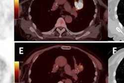
LONDON - PET/CT prior to radiotherapy can help oncologists target a more powerful dose of radiation to non-small cell lung cancer (NSCLC), whilst keeping dose low in other areas of the organ, according to research presented at this week's European Society for Therapeutic Radiology and Oncology (ESTRO) meeting.
Dutch researchers wanted to address the thorny problem of "radioresistant" tumors, or cancers that don't respond to radiation therapy delivered at conventional levels. But the concept isn't truly an accurate one, as all tumors will respond to radiation assuming the dose is high enough, according to Dr. Dirk De Ruysscher from the Maastro Clinic at University Medical Centre Maastricht.
"Radioresistance doesn't really exist in the pure sense; it's a matter of dose and timing," he said. "Stage III NSCLC is heterogeneous, with poor survival and poor local control, but even in these tumors, improving local control can improve survival."
The trick is finding a way to increase dose without affecting the organ at risk. That's where FDG-PET/CT can help by identifying areas within the tumor that might be resistant to treatment.
"By using PET, it's possible to increase radiation dose by decreasing radiation field without increased side effects," De Ruysscher said. "It is quite remarkable that this holds true for stage III NSCLC as it does for all stages."
When De Ruysscher and colleagues tested the theory in a phase II study, they irradiated lymph nodes that were positive for cancer on PET scans and monitored local recurrence. "We found only 2% isolated node failures when based on PET-positive lymph nodes," he said.
Following this, an individualized dose escalation study combined selective nodal irradiation based on FDG-PET scans with short overall treatment time based on a certain mean lung dose (MLD) up to around 90 Gy. Results, published in the Journal of Clinical Oncology, showed approximately 45% survival, and a further publication will show 55% survival with concurrent chemoradiation at two years.
Next, the group decided to investigate dose escalation when delivered "inhomogeneously." NSCLC tumors are heterogeneous in composition, so delivering equal doses across a tumor provides too low a dose to resistant parts, and overkill to more sensitive parts.
"It's more logical to give a heterogeneous dose distribution to a heterogeneous tumor for optimized local control," De Ruysscher said.
![]() |
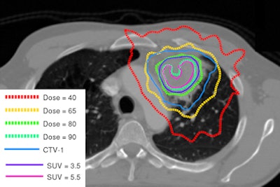
| Image shows redistribution of dose according to tumor heterogeneity. Courtesy of Dr. Dirk De Ruysscher. |
An ongoing phase II randomized trial investigating dose redistribution is being conducted with the Netherlands Cancer Institute. Instead of a flat radiation dose across the tumor (all greater than 4 cm for local tumor control), dose is increased in the FDG-positive areas. A radiation dose of 90 Gy in 24 fractions was given to some areas without subjecting normal tissues to an increased dose.
"In the inhomogeneous dose group, nodal areas received 66 Gy in 24 fractions, whilst FDG-positive areas received the highest dose possible, similar to stereotactic body radiation therapy," he said.
De Ruysscher will also investigate the value of hypoxia-related PET scans and delayed contrast-enhanced CT scans looking at blood flow, permeability, and blood volume, and correlate these metrics with FDG and hypoxia signals to understand what is happening in patients on both very high and homogeneous doses.
"We've found that hypoxic areas overlap with FDG-positive areas by up to 80%," he said.
After the presentation, an audience member questioned whether it was necessary to acquire hypoxia measurements with CT given the availability of FDG uptake information.
"A metabolic response is much better than a CT response, and that shows an important prognostic value, but I think that we need data before or early in treatment and then dose-escalate in some cases," De Ruysscher said. "We chose to image pretreatment because we believe short imaging time is important, and adaptation after three or four weeks means overall treatment time is longer."



![Examples of ultrasound findings and techniques. (A) Images in a 39-year-old male patient with a mass in the left thigh. The mass is heterogeneous on the B-mode US image (compared with the patient in D) and showed increased microvascularity (superb microvascular imaging [SMI]) and shear-wave elastography (SWE) values. Undifferentiated pleomorphic sarcoma was diagnosed at biopsy (with pleomorphic rhabdomyosarcoma in surgical specimen). (B) Images in an 18-year-old male patient with a mass in the left leg. The mass is hypoechoic on the B-mode image, with no other findings suggestive of malignancy. The lesion is in contact with the cortex of the tibia, which is slightly irregular. CT revealed a doubtful anteromedial tibial erosion. The microvascular study demonstrated high vascularization, suggestive of malignancy. Periosteal Ewing sarcoma was diagnosed with both histologic and immunohistochemical confirmation. (C) Images in a 69-year-old female patient with a lump growing on the outside of the left leg. Multiple SWE examinations were performed (please note the high values obtained in the measurements, whereas the color map highlights the stiffness relative to adjacent tissues). SMI showed areas of increased vascularization to target for sampling. Undifferentiated spindle cell sarcoma was diagnosed at biopsy, with residual leiomyosarcoma in the surgical specimen after neoadjuvant therapy. (D) Images in a 56-year-old female patient with a mass in the right thigh. The mass is heterogeneous at both B-mode ultrasound (similar to patient A) and MRI (coronal T2-weighted spectral attenuated inversion recovery [SPAIR]; T1-weighted pre-contrast and postcontrast imaging), which even shows uptake after the administration of paramagnetic contrast material, which is traditionally suggestive of malignancy. Low values at SMI and elastography are suggestive of benignity. Spindle cell lipoma was diagnosed at biopsy, with atypical spindle cell lipomatous tumor in the surgical specimen.](https://img.auntminnieeurope.com/mindful/smg/workspaces/default/uploads/2026/08/images-radiol250278fig2.APCFLSvX6p.jpg?auto=format%2Ccompress&fit=crop&h=100&q=70&w=100)


![Examples of ultrasound findings and techniques. (A) Images in a 39-year-old male patient with a mass in the left thigh. The mass is heterogeneous on the B-mode US image (compared with the patient in D) and showed increased microvascularity (superb microvascular imaging [SMI]) and shear-wave elastography (SWE) values. Undifferentiated pleomorphic sarcoma was diagnosed at biopsy (with pleomorphic rhabdomyosarcoma in surgical specimen). (B) Images in an 18-year-old male patient with a mass in the left leg. The mass is hypoechoic on the B-mode image, with no other findings suggestive of malignancy. The lesion is in contact with the cortex of the tibia, which is slightly irregular. CT revealed a doubtful anteromedial tibial erosion. The microvascular study demonstrated high vascularization, suggestive of malignancy. Periosteal Ewing sarcoma was diagnosed with both histologic and immunohistochemical confirmation. (C) Images in a 69-year-old female patient with a lump growing on the outside of the left leg. Multiple SWE examinations were performed (please note the high values obtained in the measurements, whereas the color map highlights the stiffness relative to adjacent tissues). SMI showed areas of increased vascularization to target for sampling. Undifferentiated spindle cell sarcoma was diagnosed at biopsy, with residual leiomyosarcoma in the surgical specimen after neoadjuvant therapy. (D) Images in a 56-year-old female patient with a mass in the right thigh. The mass is heterogeneous at both B-mode ultrasound (similar to patient A) and MRI (coronal T2-weighted spectral attenuated inversion recovery [SPAIR]; T1-weighted pre-contrast and postcontrast imaging), which even shows uptake after the administration of paramagnetic contrast material, which is traditionally suggestive of malignancy. Low values at SMI and elastography are suggestive of benignity. Spindle cell lipoma was diagnosed at biopsy, with atypical spindle cell lipomatous tumor in the surgical specimen.](https://img.auntminnieeurope.com/mindful/smg/workspaces/default/uploads/2026/08/images-radiol250278fig2.APCFLSvX6p.jpg?auto=format%2Ccompress&fit=crop&h=112&q=70&w=112)