Selectively increasing radiotherapy dose in tumor areas with significant uptake of the PET radiotracer F-18 fluoromisonidazole (FMISO) does not necessarily improve outcomes for non-small cell lung cancer (NSCLC) patients, according to a phase II study published in the July issue of the Journal of Nuclear Medicine.
The study was conducted by 15 academic PET facilities across France. The researchers evaluated 54 patients with localized, advanced non-small cell cancers who were undergoing chemoradiotherapy.
All patients underwent two FDG-PET/CT and two FMISO-PET/CT scans on the same scanner and under identical conditions. In 24 (70%) of 34 patients who had positive FMISO results, the radiotherapy dose was increased up to 86 Gy on hypoxic areas identified on FMISO-PET/CT. However, this additional dose did not improve patient outcomes (JNM, July 2017, Vol. 58:7, pp. 1045-1053).
At the three-month follow-up, 31 patients (57%) showed a favorable response to the radiotherapy, including 17 (50%) of the 34 patients in the FMISO-positive group. At one year, overall survival was 63%, and disease-free survival was significantly longer among patients whose FMISO scans were negative (p = 0.004). Radiotherapy dose, however, was not associated with disease-free survival when adjusted for patients' FMISO status.
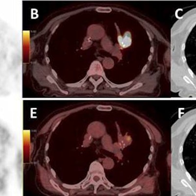
Images from a patient with an upper left lung NSCLC. (A) FDG; (B) FDG-PET/CT; (C) planning radiotherapy based on FDG (66 Gy) with BTVm (gross tumor volume), clinical target volume, and planning target volume; (D) FMISO-PET; (E) FMISO-PET/CT; and (F) boost based on the FMISO-PET (76 Gy) with BTVh (biological hypoxic target volume) and planning target volume boost. Images courtesy of Vera et al and JNM."Our approach results in a response rate of 40% or more with acceptable toxicity," concluded the group led by Dr. Pierre Vera, PhD, of the Henri Becquerel Cancer Center and Rouen University Hospital. "F-18 FMISO uptake in NSCLC patients is strongly associated with poor prognosis features that could not be reversed by radiotherapy doses up to 86 Gy."
On the positive side, the researchers noted that their approach of boosting radiotherapy based on hypoxia PET is feasible in a multicenter setting. The data also showed that smaller target volumes, identified by their functional characteristics using hypoxia PET/CT, can be adequately targeted. Finally, no significant toxicity has been associated with the radiotherapy boost.
In an invited perspective, also published in the July JNM, Dr. Rodney Hicks from the Peter MacCallum Cancer Centre and the University of Melbourne noted that negative results are instructive.
"Clearly, hypoxia remains an evil foe in our battle to achieve better outcomes in non-small cell cancer," he wrote. "But by demonstrating its importance, Vera and colleagues pose us the challenge to design new combination therapies."


![Examples of ultrasound findings and techniques. (A) Images in a 39-year-old male patient with a mass in the left thigh. The mass is heterogeneous on the B-mode US image (compared with the patient in D) and showed increased microvascularity (superb microvascular imaging [SMI]) and shear-wave elastography (SWE) values. Undifferentiated pleomorphic sarcoma was diagnosed at biopsy (with pleomorphic rhabdomyosarcoma in surgical specimen). (B) Images in an 18-year-old male patient with a mass in the left leg. The mass is hypoechoic on the B-mode image, with no other findings suggestive of malignancy. The lesion is in contact with the cortex of the tibia, which is slightly irregular. CT revealed a doubtful anteromedial tibial erosion. The microvascular study demonstrated high vascularization, suggestive of malignancy. Periosteal Ewing sarcoma was diagnosed with both histologic and immunohistochemical confirmation. (C) Images in a 69-year-old female patient with a lump growing on the outside of the left leg. Multiple SWE examinations were performed (please note the high values obtained in the measurements, whereas the color map highlights the stiffness relative to adjacent tissues). SMI showed areas of increased vascularization to target for sampling. Undifferentiated spindle cell sarcoma was diagnosed at biopsy, with residual leiomyosarcoma in the surgical specimen after neoadjuvant therapy. (D) Images in a 56-year-old female patient with a mass in the right thigh. The mass is heterogeneous at both B-mode ultrasound (similar to patient A) and MRI (coronal T2-weighted spectral attenuated inversion recovery [SPAIR]; T1-weighted pre-contrast and postcontrast imaging), which even shows uptake after the administration of paramagnetic contrast material, which is traditionally suggestive of malignancy. Low values at SMI and elastography are suggestive of benignity. Spindle cell lipoma was diagnosed at biopsy, with atypical spindle cell lipomatous tumor in the surgical specimen.](https://img.auntminnieeurope.com/mindful/smg/workspaces/default/uploads/2026/08/images-radiol250278fig2.APCFLSvX6p.jpg?auto=format%2Ccompress&fit=crop&h=100&q=70&w=100)


![Examples of ultrasound findings and techniques. (A) Images in a 39-year-old male patient with a mass in the left thigh. The mass is heterogeneous on the B-mode US image (compared with the patient in D) and showed increased microvascularity (superb microvascular imaging [SMI]) and shear-wave elastography (SWE) values. Undifferentiated pleomorphic sarcoma was diagnosed at biopsy (with pleomorphic rhabdomyosarcoma in surgical specimen). (B) Images in an 18-year-old male patient with a mass in the left leg. The mass is hypoechoic on the B-mode image, with no other findings suggestive of malignancy. The lesion is in contact with the cortex of the tibia, which is slightly irregular. CT revealed a doubtful anteromedial tibial erosion. The microvascular study demonstrated high vascularization, suggestive of malignancy. Periosteal Ewing sarcoma was diagnosed with both histologic and immunohistochemical confirmation. (C) Images in a 69-year-old female patient with a lump growing on the outside of the left leg. Multiple SWE examinations were performed (please note the high values obtained in the measurements, whereas the color map highlights the stiffness relative to adjacent tissues). SMI showed areas of increased vascularization to target for sampling. Undifferentiated spindle cell sarcoma was diagnosed at biopsy, with residual leiomyosarcoma in the surgical specimen after neoadjuvant therapy. (D) Images in a 56-year-old female patient with a mass in the right thigh. The mass is heterogeneous at both B-mode ultrasound (similar to patient A) and MRI (coronal T2-weighted spectral attenuated inversion recovery [SPAIR]; T1-weighted pre-contrast and postcontrast imaging), which even shows uptake after the administration of paramagnetic contrast material, which is traditionally suggestive of malignancy. Low values at SMI and elastography are suggestive of benignity. Spindle cell lipoma was diagnosed at biopsy, with atypical spindle cell lipomatous tumor in the surgical specimen.](https://img.auntminnieeurope.com/mindful/smg/workspaces/default/uploads/2026/08/images-radiol250278fig2.APCFLSvX6p.jpg?auto=format%2Ccompress&fit=crop&h=112&q=70&w=112)