Dear AuntMinnieEurope Member,
If you were to ask Dr. Thomas Vogl which imaging modality excites him most, the answer might well come as a surprise. He is well known for his MR research, but he seems even more passionate these days about the clinical applications and future potential of digital x-ray.
Sustained progress is being made in digital radiography, as well as computed radiography. Direct detectors represent a small but spectacular revolution, according to Vogl and his research group in Frankfurt am Main, Germany, who are working hard to advance this revolution. As deputy editor of European Radiology, he is used to selecting his words carefully, so it's significant that he refers to these detectors in such terms.
You can read about these and other developments in our new Digital X-Ray Community. To coincide with the launch of this important section, we have published two articles involving the team in Frankfurt, including an exclusive interview with Vogl himself. Click here to find out more, or visit the community directly at xray.auntminnieeurope.com.
The football season is reaching its annual climax across Europe, and the sport has even made it into the hallowed pages of the Lancet. Its latest edition contains fascinating details of a freak accident involving a 15-year-old player from Munich. To read how CT provided vital presurgical information, click here.
We're also entering a hectic period in the congress season. We have two conferences going on this week: the annual meeting of the International Society for Magnetic Resonance in Medicine (ISMRM) in Montreal, Canada, and the European Society for Therapeutic Radiology and Oncology (ESTRO) in London.
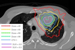
From the ISMRM show, you can click here to read our first news report from the event, on the use of a new blood-pool contrast agent for MR angiography. From ESTRO, we have a report on using PET/CT to direct more targeted radiation therapy of non-small cell lung cancer tumors. Click here for that story.
We're committed to bringing you news from all the major global meetings, and over the next few days watch out for coverage of the World of Health IT conference in Budapest in our Healthcare Informatics Digital Community.
If you haven't done so already, take a look at our Forums, which have been alive with activity this month. Dr. Peter Rinck's recent column about role extension of radiographers has generated much discussion, and you can join the debate by clicking here.


![Examples of ultrasound findings and techniques. (A) Images in a 39-year-old male patient with a mass in the left thigh. The mass is heterogeneous on the B-mode US image (compared with the patient in D) and showed increased microvascularity (superb microvascular imaging [SMI]) and shear-wave elastography (SWE) values. Undifferentiated pleomorphic sarcoma was diagnosed at biopsy (with pleomorphic rhabdomyosarcoma in surgical specimen). (B) Images in an 18-year-old male patient with a mass in the left leg. The mass is hypoechoic on the B-mode image, with no other findings suggestive of malignancy. The lesion is in contact with the cortex of the tibia, which is slightly irregular. CT revealed a doubtful anteromedial tibial erosion. The microvascular study demonstrated high vascularization, suggestive of malignancy. Periosteal Ewing sarcoma was diagnosed with both histologic and immunohistochemical confirmation. (C) Images in a 69-year-old female patient with a lump growing on the outside of the left leg. Multiple SWE examinations were performed (please note the high values obtained in the measurements, whereas the color map highlights the stiffness relative to adjacent tissues). SMI showed areas of increased vascularization to target for sampling. Undifferentiated spindle cell sarcoma was diagnosed at biopsy, with residual leiomyosarcoma in the surgical specimen after neoadjuvant therapy. (D) Images in a 56-year-old female patient with a mass in the right thigh. The mass is heterogeneous at both B-mode ultrasound (similar to patient A) and MRI (coronal T2-weighted spectral attenuated inversion recovery [SPAIR]; T1-weighted pre-contrast and postcontrast imaging), which even shows uptake after the administration of paramagnetic contrast material, which is traditionally suggestive of malignancy. Low values at SMI and elastography are suggestive of benignity. Spindle cell lipoma was diagnosed at biopsy, with atypical spindle cell lipomatous tumor in the surgical specimen.](https://img.auntminnieeurope.com/mindful/smg/workspaces/default/uploads/2026/08/images-radiol250278fig2.APCFLSvX6p.jpg?auto=format%2Ccompress&fit=crop&h=100&q=70&w=100)


![Examples of ultrasound findings and techniques. (A) Images in a 39-year-old male patient with a mass in the left thigh. The mass is heterogeneous on the B-mode US image (compared with the patient in D) and showed increased microvascularity (superb microvascular imaging [SMI]) and shear-wave elastography (SWE) values. Undifferentiated pleomorphic sarcoma was diagnosed at biopsy (with pleomorphic rhabdomyosarcoma in surgical specimen). (B) Images in an 18-year-old male patient with a mass in the left leg. The mass is hypoechoic on the B-mode image, with no other findings suggestive of malignancy. The lesion is in contact with the cortex of the tibia, which is slightly irregular. CT revealed a doubtful anteromedial tibial erosion. The microvascular study demonstrated high vascularization, suggestive of malignancy. Periosteal Ewing sarcoma was diagnosed with both histologic and immunohistochemical confirmation. (C) Images in a 69-year-old female patient with a lump growing on the outside of the left leg. Multiple SWE examinations were performed (please note the high values obtained in the measurements, whereas the color map highlights the stiffness relative to adjacent tissues). SMI showed areas of increased vascularization to target for sampling. Undifferentiated spindle cell sarcoma was diagnosed at biopsy, with residual leiomyosarcoma in the surgical specimen after neoadjuvant therapy. (D) Images in a 56-year-old female patient with a mass in the right thigh. The mass is heterogeneous at both B-mode ultrasound (similar to patient A) and MRI (coronal T2-weighted spectral attenuated inversion recovery [SPAIR]; T1-weighted pre-contrast and postcontrast imaging), which even shows uptake after the administration of paramagnetic contrast material, which is traditionally suggestive of malignancy. Low values at SMI and elastography are suggestive of benignity. Spindle cell lipoma was diagnosed at biopsy, with atypical spindle cell lipomatous tumor in the surgical specimen.](https://img.auntminnieeurope.com/mindful/smg/workspaces/default/uploads/2026/08/images-radiol250278fig2.APCFLSvX6p.jpg?auto=format%2Ccompress&fit=crop&h=112&q=70&w=112)