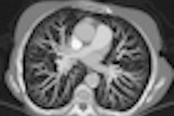
Computer-aided detection (CAD) can significantly improve the sensitivity of pulmonary embolism (PE) detection among inexperienced readers, and despite a slight increase in the number of false positives, the added value of CAD is particularly evident on a patient-based analysis.
These are among the key findings of a study published in the June print edition of European Radiology (Vol. 21:12, pp.1214-1223).
Improved PE detection may lead to accurate classification of patients in on-call situations; for instance, where reading time and the lack of experienced staff are often critical factors. Increases in sensitivity and specificity are desirable to enhance the clinical use of CAD algorithms and optimize patient management, according to lead author Dr. Kevin Blackmon, from the department of radiology and radiological science at the Medical University of South Carolina.
"CAD improves detection of lesions while avoiding the labor burden of a double or consensus read," he noted. "Unlike human observer performance that can vary with fatigue, time constraints, or experience level, CAD performance is constant and the level of performance is intrinsic to the sophistication of the algorithm."
To evaluate the effect of CAD on the performance of novices for PE detection using CT pulmonary angiography (CTPA), the researchers analyzed the examinations of 79 patients. The patients were between 18 and 52 years of age, and 50 of them were women. A 16-slice CT unit (Somatom Sensation 16, Siemens Healthcare, or LightSpeed, GE Healthcare) was used for 35 exams, while a 64-slice CT system (Somatom Sensation 64, Siemens) was used for 42 exams. A dual-source CT (Somatom Definition, Siemens) was used in the remaining two cases.
Studies were evaluated by two independent inexperienced readers who marked all vessels containing PE. After three months, the studies were re-evaluated by the same two readers, this time aided by a CAD prototype (VA10 PE, Siemens).
A consensus read by three expert radiologists revealed 119 PEs in 32 of the 79 patients. For PE detection, the sensitivity of CAD alone was 78%. Inexperienced readers' initial interpretations had an average per-PE sensitivity of 50%, which improved to 71% (p < 0.001) with CAD as a second reader. False positives increased from 0.18 to 0.25 per study (p = 0.03). The readers initially detected 27 out of 32 positive studies (84%), but with CAD this number increased to 29.5 studies (92%, p = 0.125).
The research has some limitations, however. Due to the retrospective nature of the study, the potential for selection bias exists, although the authors attempted to minimize this by including consecutive positive and negative studies from the same start date. Another limitation was the use of CAD during the expert consensus read to establish the presence and location of PE. This could have led to ambiguous lesions being called PE.
Furthermore, the population size was small and there is potential for recall bias because the same cohort of cases was evaluated with and without CAD, although measures were taken to avoid this bias. Because of a dwindling number of positive PE cases at CTPA in the authors' institutions, the population was enriched with 32 patients whose datasets were initially interpreted as positive for PE before being included in the study.
A strength of the research was the analysis of emboli at all vessel levels. Additionally, considering that PE diagnosis ultimately occurs at a patient level, the use of CAD enabled improvement of the detection rate of patients with confirmed PE, significantly increasing the per-study sensitivity.
Future studies will look at the overall effect of PE CAD on patient management, which will be essential to widespread adoption of the technology, Blackmon noted. The researchers also plan to study the potential integration of other quantifiable clinical factors, such as overall clot-burden and quantification of right ventricular strain to determine optimal patient management.


![Examples of ultrasound findings and techniques. (A) Images in a 39-year-old male patient with a mass in the left thigh. The mass is heterogeneous on the B-mode US image (compared with the patient in D) and showed increased microvascularity (superb microvascular imaging [SMI]) and shear-wave elastography (SWE) values. Undifferentiated pleomorphic sarcoma was diagnosed at biopsy (with pleomorphic rhabdomyosarcoma in surgical specimen). (B) Images in an 18-year-old male patient with a mass in the left leg. The mass is hypoechoic on the B-mode image, with no other findings suggestive of malignancy. The lesion is in contact with the cortex of the tibia, which is slightly irregular. CT revealed a doubtful anteromedial tibial erosion. The microvascular study demonstrated high vascularization, suggestive of malignancy. Periosteal Ewing sarcoma was diagnosed with both histologic and immunohistochemical confirmation. (C) Images in a 69-year-old female patient with a lump growing on the outside of the left leg. Multiple SWE examinations were performed (please note the high values obtained in the measurements, whereas the color map highlights the stiffness relative to adjacent tissues). SMI showed areas of increased vascularization to target for sampling. Undifferentiated spindle cell sarcoma was diagnosed at biopsy, with residual leiomyosarcoma in the surgical specimen after neoadjuvant therapy. (D) Images in a 56-year-old female patient with a mass in the right thigh. The mass is heterogeneous at both B-mode ultrasound (similar to patient A) and MRI (coronal T2-weighted spectral attenuated inversion recovery [SPAIR]; T1-weighted pre-contrast and postcontrast imaging), which even shows uptake after the administration of paramagnetic contrast material, which is traditionally suggestive of malignancy. Low values at SMI and elastography are suggestive of benignity. Spindle cell lipoma was diagnosed at biopsy, with atypical spindle cell lipomatous tumor in the surgical specimen.](https://img.auntminnieeurope.com/mindful/smg/workspaces/default/uploads/2026/08/images-radiol250278fig2.APCFLSvX6p.jpg?auto=format%2Ccompress&fit=crop&h=100&q=70&w=100)


![Examples of ultrasound findings and techniques. (A) Images in a 39-year-old male patient with a mass in the left thigh. The mass is heterogeneous on the B-mode US image (compared with the patient in D) and showed increased microvascularity (superb microvascular imaging [SMI]) and shear-wave elastography (SWE) values. Undifferentiated pleomorphic sarcoma was diagnosed at biopsy (with pleomorphic rhabdomyosarcoma in surgical specimen). (B) Images in an 18-year-old male patient with a mass in the left leg. The mass is hypoechoic on the B-mode image, with no other findings suggestive of malignancy. The lesion is in contact with the cortex of the tibia, which is slightly irregular. CT revealed a doubtful anteromedial tibial erosion. The microvascular study demonstrated high vascularization, suggestive of malignancy. Periosteal Ewing sarcoma was diagnosed with both histologic and immunohistochemical confirmation. (C) Images in a 69-year-old female patient with a lump growing on the outside of the left leg. Multiple SWE examinations were performed (please note the high values obtained in the measurements, whereas the color map highlights the stiffness relative to adjacent tissues). SMI showed areas of increased vascularization to target for sampling. Undifferentiated spindle cell sarcoma was diagnosed at biopsy, with residual leiomyosarcoma in the surgical specimen after neoadjuvant therapy. (D) Images in a 56-year-old female patient with a mass in the right thigh. The mass is heterogeneous at both B-mode ultrasound (similar to patient A) and MRI (coronal T2-weighted spectral attenuated inversion recovery [SPAIR]; T1-weighted pre-contrast and postcontrast imaging), which even shows uptake after the administration of paramagnetic contrast material, which is traditionally suggestive of malignancy. Low values at SMI and elastography are suggestive of benignity. Spindle cell lipoma was diagnosed at biopsy, with atypical spindle cell lipomatous tumor in the surgical specimen.](https://img.auntminnieeurope.com/mindful/smg/workspaces/default/uploads/2026/08/images-radiol250278fig2.APCFLSvX6p.jpg?auto=format%2Ccompress&fit=crop&h=112&q=70&w=112)