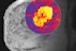
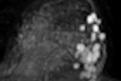
When trying to find ways to cut contrast in patients scheduled for breast MRI, Italian researchers discovered that a noncontrast water-fat separation sequence was no better for evaluating tumor response to neoadjuvant chemotherapy than standard contrast MRI. But noncontrast MRI still holds potential for monitoring therapy.
The group from the University of Rome "La Sapienza" concluded that apparent diffusion coefficient (ADC) values may be useful in differentiating patients who respond well to early chemotherapy from those who do not respond adequately to therapy.
"Nowadays, contrast-enhanced MRI is commonly used in many centers as a diagnostic tool in monitoring neoadjuvant chemotherapy response in patients affected by breast cancer," said lead study author Dr. Federica Pediconi in her presentation at the 2011 European Congress of Radiology (ECR). "However, contrast agents may increase examination time and cost, and may lead to different media contrast reactions" such as nephrogenic systemic fibrosis in patients with impaired renal function.
Given the potential of adverse reactions to MRI contrast agents, Pediconi and colleagues utilized diffusion-weighted imaging (DWI) without contrast in combination with a T2-weighted protocol called iterative decomposition of water and fat with echo asymmetry and least-squares estimation (IDEAL).
IDEAL is a water-fat separation technique that can be applied to a variety of MR pulse sequences, she explained. One advantage of IDEAL imaging is that fat-only and water-only MR images can be obtained during a single scan. Previous research has shown that DWI could help detect breast lesions and aid in the early assessment of treatment response in breast cancer patients.
Pilot study purpose
The goal of the pilot study was to determine the sensitivity and specificity of 3-tesla MRI using DWI T2-weighted IDEAL images and no contrast agent, and compare those results with contrast-enhanced MRI to assess tumor response to neoadjuvant chemotherapy.
Twelve consecutive patients underwent neoadjuvant chemotherapy for biopsy-proven breast cancer between January 2010 and May 2010. The breast lesions were larger than 2 cm. The patients also received 3-tesla MRI scans (Signa HDx, GE Healthcare) prior to chemotherapy and before surgical excision.
There were 11 patients with invasive ductal carcinoma: seven completely responded to chemotherapy, two partially responded, and two had no response. The remaining patient had an invasive lobular carcinoma and responded completely to chemotherapy.
MRI protocol
The MRI protocol included fast spin-echo T2-weighted IDEAL and DWI sequences, and a 3D T1-weighted sequence acquired before and after administration of gadobenate dimeglumine (MultiHance, Bracco Diagnostics).
The researchers evaluated tumor response in the MR images after chemotherapy and compared the results with histological specimens obtained after surgery. Two observers also rated the images from T2-weighted studies and images from contrast-enhanced MRI to evaluate tumor response.
In a comparison of the two readers' results, the first observer achieved sensitivity of 92% for T2-weighted IDEAL MR images, while the second reader achieved sensitivity of 84%. Both readers achieved sensitivity of 100% for contrast-enhanced MRI. Similar specificity results were noted by the two observers: 82% for T2-weighted IDEAL images and 86% for contrast-enhanced MRI.
The differences between methods and between observers for sensitivity and specificity were not statistically significant, Pediconi and colleagues noted. In addition, no significant differences in lesion size measurements were found between sequences.
"Although we didn't find a statistically significant difference between contrast-enhanced MRI and unenhanced MRI, the visibility of the lesions was better by using contrast," she said.
ADC values
The researchers also compared patients' ADC values at baseline and after two cycles of neoadjuvant chemotherapy to see how well they responded to the treatment. The mean ADC value prior to chemotherapy was 1.32, while the ADC value after neoadjuvant chemotherapy was 1.59.
"The meaning of the ADC values is that if we find an increasing value of ADC after two cycles, we can predict a good response to the treatment," Pediconi said. "So, the clinicians can use this factor to distinguish responders and nonresponders."
Based on the results, the "combination of diffusion-weighted imaging for tumor density and T2-weighted IDEAL imaging for lesion morphology and size is similar to contrast-enhanced MRI for the evaluation of tumor response to neoadjuvant chemotherapy," the researchers concluded.
Given the findings, physicians may be able to "monitor this patient group without contrast agents in the future." However, they added that contrast-enhanced MRI still outperformed the other two MRI techniques for sensitivity and specificity.