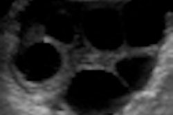
Transvaginal ultrasound screening can detect endometrial cancer before symptoms appear in postmenopausal women, according to research from the U.K. published online in Lancet Oncology.
In a large-scale study to assess the performance of transvaginal ultrasound screening for endometrial cancer, a research team led by Ian Jacobs, of University College London, found that the modality yielded 81% sensitivity and 86% specificity.
The researchers analyzed data from the United Kingdom Collaborative Trial of Ovarian Cancer Screening (UKCTOCS), in which transvaginal ultrasound screening was performed on 37,038 women (Lancet Oncology, December 13, 2010).
Performance characteristics of endometrial thickness and endometrial abnormalities at detecting endometrial cancer within one year of transvaginal ultrasound screening were calculated. The researchers then evaluated the method's sensitivity and specificity for different risk groups, and the group performed modeling using epidemiological variables to assess a screening strategy for women at higher risk.
Of the 133 women diagnosed with endometrial cancer or atypical endometrial hyperplasia within one year of screening, 107 (81%) had an endometrial thickness of 5 mm or more. Most of the 36,731 women who did not have cancer had an endometrial thickness of less than 5 mm, according to the researchers.
At the optimum cutoff of 5.15 mm, transvaginal ultrasound would detect 80.5% of cancer cases, with a false-positive rate of 14.3% (85.7% specificity), they found. With this screening threshold, one case of endometrial cancer would be detected for every 47.7 women screened, according to the researchers.
The authors noted, however, that further studies are needed to examine transvaginal ultrasound screening's acceptability, health economics, and risk before routine population screening or targeted screening can be advocated.
By Erik L. Ridley
AuntMinnie.com staff writer
December 13, 2010
Related Reading
Hysteroscopy still beats sonography for diagnosing endometrial pathology, June 3, 2010
Ultrasound superior to symptoms for predicting ovarian cancer, July 16, 2009
Transvaginal ultrasound screening fails to detect early-stage ovarian cancer, December 26, 2007
Copyright © 2010 AuntMinnie.com


![Examples of ultrasound findings and techniques. (A) Images in a 39-year-old male patient with a mass in the left thigh. The mass is heterogeneous on the B-mode US image (compared with the patient in D) and showed increased microvascularity (superb microvascular imaging [SMI]) and shear-wave elastography (SWE) values. Undifferentiated pleomorphic sarcoma was diagnosed at biopsy (with pleomorphic rhabdomyosarcoma in surgical specimen). (B) Images in an 18-year-old male patient with a mass in the left leg. The mass is hypoechoic on the B-mode image, with no other findings suggestive of malignancy. The lesion is in contact with the cortex of the tibia, which is slightly irregular. CT revealed a doubtful anteromedial tibial erosion. The microvascular study demonstrated high vascularization, suggestive of malignancy. Periosteal Ewing sarcoma was diagnosed with both histologic and immunohistochemical confirmation. (C) Images in a 69-year-old female patient with a lump growing on the outside of the left leg. Multiple SWE examinations were performed (please note the high values obtained in the measurements, whereas the color map highlights the stiffness relative to adjacent tissues). SMI showed areas of increased vascularization to target for sampling. Undifferentiated spindle cell sarcoma was diagnosed at biopsy, with residual leiomyosarcoma in the surgical specimen after neoadjuvant therapy. (D) Images in a 56-year-old female patient with a mass in the right thigh. The mass is heterogeneous at both B-mode ultrasound (similar to patient A) and MRI (coronal T2-weighted spectral attenuated inversion recovery [SPAIR]; T1-weighted pre-contrast and postcontrast imaging), which even shows uptake after the administration of paramagnetic contrast material, which is traditionally suggestive of malignancy. Low values at SMI and elastography are suggestive of benignity. Spindle cell lipoma was diagnosed at biopsy, with atypical spindle cell lipomatous tumor in the surgical specimen.](https://img.auntminnieeurope.com/mindful/smg/workspaces/default/uploads/2026/08/images-radiol250278fig2.APCFLSvX6p.jpg?auto=format%2Ccompress&fit=crop&h=100&q=70&w=100)


![Examples of ultrasound findings and techniques. (A) Images in a 39-year-old male patient with a mass in the left thigh. The mass is heterogeneous on the B-mode US image (compared with the patient in D) and showed increased microvascularity (superb microvascular imaging [SMI]) and shear-wave elastography (SWE) values. Undifferentiated pleomorphic sarcoma was diagnosed at biopsy (with pleomorphic rhabdomyosarcoma in surgical specimen). (B) Images in an 18-year-old male patient with a mass in the left leg. The mass is hypoechoic on the B-mode image, with no other findings suggestive of malignancy. The lesion is in contact with the cortex of the tibia, which is slightly irregular. CT revealed a doubtful anteromedial tibial erosion. The microvascular study demonstrated high vascularization, suggestive of malignancy. Periosteal Ewing sarcoma was diagnosed with both histologic and immunohistochemical confirmation. (C) Images in a 69-year-old female patient with a lump growing on the outside of the left leg. Multiple SWE examinations were performed (please note the high values obtained in the measurements, whereas the color map highlights the stiffness relative to adjacent tissues). SMI showed areas of increased vascularization to target for sampling. Undifferentiated spindle cell sarcoma was diagnosed at biopsy, with residual leiomyosarcoma in the surgical specimen after neoadjuvant therapy. (D) Images in a 56-year-old female patient with a mass in the right thigh. The mass is heterogeneous at both B-mode ultrasound (similar to patient A) and MRI (coronal T2-weighted spectral attenuated inversion recovery [SPAIR]; T1-weighted pre-contrast and postcontrast imaging), which even shows uptake after the administration of paramagnetic contrast material, which is traditionally suggestive of malignancy. Low values at SMI and elastography are suggestive of benignity. Spindle cell lipoma was diagnosed at biopsy, with atypical spindle cell lipomatous tumor in the surgical specimen.](https://img.auntminnieeurope.com/mindful/smg/workspaces/default/uploads/2026/08/images-radiol250278fig2.APCFLSvX6p.jpg?auto=format%2Ccompress&fit=crop&h=112&q=70&w=112)