A prototype computer-aided detection (CAD) algorithm for tuberculosis (TB) developed by Dutch researchers performed well in a pilot study, offering hope that the software could enable the use of chest digital radiography (DR) to screen for TB in areas with a high burden of the disease but not many skilled readers.
A research team led by Laurens Hogeweg, PhD, from Radboud University Nijmegen Medical Center in the Netherlands, reported promising initial results from prototype CAD software that was compared to an expert human reader for evaluating TB. While the software is still being developed, the researchers believe that the technology may one day help make up for the lack of necessary radiologists in countries with large numbers of TB cases.
Hogeweg shared the research -- a joint venture between the University of Nijmegen, the Zambia AIDS Related Tuberculosis (ZAMBART) project, and the University of Cape Town in South Africa -- during a scientific session at the 2010 RSNA meeting in Chicago.
TB is a major healthcare problem, with up to one-third of the world's population infected, according to the World Health Organization (WHO). There are 9 million new infections and 2 million deaths each year, Hogeweg said.
While a cheap and effective cure for TB is available, it's essential to detect active TB cases, Hogeweg said. Digital chest x-rays have been proposed for this purpose, but there are few skilled chest x-ray readers in many developing countries with a high incidence of TB and few healthcare resources.
"For example, in Zambia there's only one radiologist available for the whole country," he said. "In such settings, CAD as an extra reader can really make a difference."
To evaluate the performance of a CAD algorithm developed at the university to analyze signs of TB on chest radiographs, the researchers used 425 digital chest radiographs from an Odelca-DR system (Delft Imaging Systems, Veenendaal, the Netherlands) with a 0.25-mm pixel size. Assessment by an expert chest radiologist served as the reference standard.
A training set for the CAD software was provided by the University of Cape Town Lung Institute in South Africa, which contributed 216 images (110 normal and 106 abnormal studies).
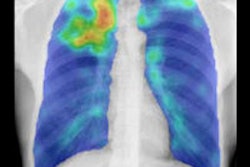
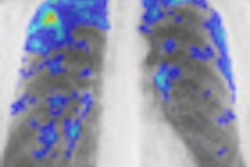
The CAD software automatically analyzes chest x-rays, detects abnormalities, and indicates the likelihood that these abnormal signs are consistent with TB and with active TB, according to Hogeweg.
The software aims to detect diffuse abnormalities via textural abnormality analysis and lung shape distortions using shape analysis. Probabilities from the textural system and the shape system are then averaged to obtain a final probability of being abnormal.
For the accurate diagnosis of normal or abnormal, the combination of texture abnormality and shape distortion analysis yielded an area under the receiver operator characteristics (ROC) curve of 0.81, when the expert reader's performance was considered as the reference standard. Texture and shape analysis alone led to an area under the curve of 0.76 and 0.80, respectively.
In future work, the researchers would like to add to the CAD algorithm the ability to analyze other morphological characteristics that could be signs of TB. While the algorithm is improved, the group is also planning a number of larger observer and clinical studies to further evaluate its performance.
"In the future, the software will be bundled with clinical x-ray machines used in the field in several countries in southern Africa," Hogeweg told AuntMinnie.com. "The software can then play a role in allowing large-scale tuberculosis screening programs to be run by reducing the number of images that have to be read."
The software could also be deployed in daily clinical practice, he said.
"The opinion of the software could be presented to a clinical officer, allowing a diagnosis of TB on the spot, and a TB suspect could immediately be put on treatment," he said.
By Erik L. Ridley
AuntMinnie.com staff writer
January 5, 2011
Related Reading
iPad holds up well for assessing tuberculosis, November 29, 2010
Bundling of CAD software boosts lung lesion detection at CT, September 21, 2010
Chest x-ray CAD offers value in detecting lung cancers, June 18, 2010
Low-dose tomo beats chest DR for TB evaluations, March 19, 2010
WHITIA targets lung disease with remote x-ray unit, November 27, 2009
Copyright © 2011 AuntMinnie.com


