VIENNA - A German research group presented preliminary work on a dedicated breast CT scanner at this week's European Congress of Radiology (ECR). While the group has only studied phantoms with the prototype system, their results so far indicate that the scanner can achieve twice the resolution of conventional mammography at an equivalent radiation dose.
CT has much higher image resolution than radiography-based techniques like mammography, which could be a boon in trying to detect the small structures that can be signs of early-stage breast cancer. But CT's radiation dose is typically much higher than that of mammography, which could be a major issue due to the need to conduct repeat studies if used in a screening environment.
No dedicated breast CT system is on the market as of yet, but groups working to develop commercial systems include the University of California, Davis, and the University of Rochester in New York, which has licensed its technology to Koning of West Henrietta, NY.
In a session on new technologies on Saturday afternoon, Sabrina Vollmar from the Institute of Medical Physics at Friedrich-Alexander-University in Erlangen-Nuremberg, Germany, discussed the university's approach to breast CT. Working as part of a group that includes CT pioneer Willi Kalender, Ph.D., university researchers have built a prototype unit in which a woman lies prone on the patient table, with the breast hanging pendant-style through a hole in the table. The system is based on conebeam CT geometry, and does not use breast compression, unlike conventional mammography.
The system features a 20 x 20-cm field-of-view, and acquires 360 projection images per gantry rotation. The system has a pixel size of 200 microns. This compares to two projections typically used in mammography, with full-field digital mammography systems typically having pixel sizes of 100 microns, Vollmar said.
To study the breast CT system's imaging characteristics in comparison to mammography, the group designed two phantoms, one for use with each modality. The phantoms were of different sizes to simulate the size of the breast when imaged with the different techniques, with the breast CT phantom measuring 10 x 10 cm while the second one was 5 x 14 cm. The phantoms included structures simulating both soft-tissue tumors and microcalcifications.
To measure radiation dose for each modality, the group used the Monte Carlo method, and measured 3D dose distribution across the entire phantom. They also measured the integral dose for both phantoms. To assess image quality the group calculated contrast-to-noise ratio (CNR) and dose-weighted contrast-to-noise ratio (CNRD).
The group imaged the phantoms at energies ranging from 15 keV to 100 keV, with the group finding that the best energy ranges for breast CT in terms of CNR was 28 keV, and for CNRD at 34 keV. Dose increased at higher energies used, Vollmar said.
Both systems delivered comparable radiation dose to the phantoms. The breast CT system delivered 4.5 mGy per study, versus 4.8 mGy for mammography when both projections from a standard two-projection study were added together.
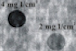
As for image quality, Vollmar said that both systems performed comparably in the detection of microcalcifications. But for tumors, the breast CT unit had twice the image quality of mammography -- the system was able to detect "tumors" in the phantom as small as 2 mm, versus 4 mm for the mammography system.
"Breast CT offers the potential for the detection of small soft-tissue lesions, makes compression of the breast obsolete, and offers image quality at least equivalent to mammography, without an increase of dose level," Vollmar said.
How soon could the group move from a prototype system to a unit able to scan patients? In remarks after Vollmar's presentation, Kalender estimated that such a system could be a couple years away.
By Brian Casey
AuntMinnie.com staff writer
March 11, 2007
Related Reading
Koning introduces CT for breast imaging, November 26, 2006
Imaging start-up to install conebeam CT breast imaging prototype, May 1, 2006
Breast CT developers aim to 'revolutionize' mammography, December 20, 2005
Researchers make inroads with breast CT, gear up for clinical test, July 25, 2005
UC Davis tests breast CT, May 11, 2005
Copyright © 2007 AuntMinnie.com


![Examples of ultrasound findings and techniques. (A) Images in a 39-year-old male patient with a mass in the left thigh. The mass is heterogeneous on the B-mode US image (compared with the patient in D) and showed increased microvascularity (superb microvascular imaging [SMI]) and shear-wave elastography (SWE) values. Undifferentiated pleomorphic sarcoma was diagnosed at biopsy (with pleomorphic rhabdomyosarcoma in surgical specimen). (B) Images in an 18-year-old male patient with a mass in the left leg. The mass is hypoechoic on the B-mode image, with no other findings suggestive of malignancy. The lesion is in contact with the cortex of the tibia, which is slightly irregular. CT revealed a doubtful anteromedial tibial erosion. The microvascular study demonstrated high vascularization, suggestive of malignancy. Periosteal Ewing sarcoma was diagnosed with both histologic and immunohistochemical confirmation. (C) Images in a 69-year-old female patient with a lump growing on the outside of the left leg. Multiple SWE examinations were performed (please note the high values obtained in the measurements, whereas the color map highlights the stiffness relative to adjacent tissues). SMI showed areas of increased vascularization to target for sampling. Undifferentiated spindle cell sarcoma was diagnosed at biopsy, with residual leiomyosarcoma in the surgical specimen after neoadjuvant therapy. (D) Images in a 56-year-old female patient with a mass in the right thigh. The mass is heterogeneous at both B-mode ultrasound (similar to patient A) and MRI (coronal T2-weighted spectral attenuated inversion recovery [SPAIR]; T1-weighted pre-contrast and postcontrast imaging), which even shows uptake after the administration of paramagnetic contrast material, which is traditionally suggestive of malignancy. Low values at SMI and elastography are suggestive of benignity. Spindle cell lipoma was diagnosed at biopsy, with atypical spindle cell lipomatous tumor in the surgical specimen.](https://img.auntminnieeurope.com/mindful/smg/workspaces/default/uploads/2026/08/images-radiol250278fig2.APCFLSvX6p.jpg?auto=format%2Ccompress&fit=crop&h=100&q=70&w=100)


![Examples of ultrasound findings and techniques. (A) Images in a 39-year-old male patient with a mass in the left thigh. The mass is heterogeneous on the B-mode US image (compared with the patient in D) and showed increased microvascularity (superb microvascular imaging [SMI]) and shear-wave elastography (SWE) values. Undifferentiated pleomorphic sarcoma was diagnosed at biopsy (with pleomorphic rhabdomyosarcoma in surgical specimen). (B) Images in an 18-year-old male patient with a mass in the left leg. The mass is hypoechoic on the B-mode image, with no other findings suggestive of malignancy. The lesion is in contact with the cortex of the tibia, which is slightly irregular. CT revealed a doubtful anteromedial tibial erosion. The microvascular study demonstrated high vascularization, suggestive of malignancy. Periosteal Ewing sarcoma was diagnosed with both histologic and immunohistochemical confirmation. (C) Images in a 69-year-old female patient with a lump growing on the outside of the left leg. Multiple SWE examinations were performed (please note the high values obtained in the measurements, whereas the color map highlights the stiffness relative to adjacent tissues). SMI showed areas of increased vascularization to target for sampling. Undifferentiated spindle cell sarcoma was diagnosed at biopsy, with residual leiomyosarcoma in the surgical specimen after neoadjuvant therapy. (D) Images in a 56-year-old female patient with a mass in the right thigh. The mass is heterogeneous at both B-mode ultrasound (similar to patient A) and MRI (coronal T2-weighted spectral attenuated inversion recovery [SPAIR]; T1-weighted pre-contrast and postcontrast imaging), which even shows uptake after the administration of paramagnetic contrast material, which is traditionally suggestive of malignancy. Low values at SMI and elastography are suggestive of benignity. Spindle cell lipoma was diagnosed at biopsy, with atypical spindle cell lipomatous tumor in the surgical specimen.](https://img.auntminnieeurope.com/mindful/smg/workspaces/default/uploads/2026/08/images-radiol250278fig2.APCFLSvX6p.jpg?auto=format%2Ccompress&fit=crop&h=112&q=70&w=112)