VIENNA - If the results of a phantom study are valid in patients, the choice of reconstruction kernel has a critical impact on coronary artery stent evaluations. And as Dr. Florian Wolf explained at today's cardiac imaging sessions at the European Congress of Radiology (ECR), the evaluation of stents for detecting restenosis is critical for patient outcomes.
"The number of implanted coronary artery stents has increased dramatically in recent years. For example in Europe in 2002, up to 600,000 coronary artery stents were implanted," said Wolf, who is from the Medical University of Vienna in Austria.
With a restenosis rate as high as 40%, there is a continuous need to evaluate implanted devices, though it can be reduced considerably with the use of drug-eluting stents.
"Despite all advances in stent technology, early detection of restenosis is crucial to improve long-term clinical outcomes and avoid recurrent ischemic periods and the occurrence of myocardial infarction," Wolf said, adding that the gold standard heart imaging modality, invasive coronary angiography, is not ideal for stent evaluation, leaving much of the work to multidetector CT.
"The purpose of our study was to evaluate whether the use of different reconstruction kernels has a significant influence on the assessment of coronary artery stents with a 64-slice CT scanner," Wolf said.
The study by Wolf, Dr. Harald Homolka, Dr. Jürgen Langenberger, Dr. Christian Herold, and colleagues examined 12 different coronary artery stents with outer diameters ranging between 2.6 mm and 5 mm. The stents were placed in a heart phantom made of a muscle-equivalent plastic material that was optimized for CT scanning.
The stents were placed into holes drilled into the phantom to simulate coronary arteries, and filled with contrast media, diluted to a CT attenuation of 300 HU. The phantom was scanned on a 64-detector-row scanner (Siemens Sensation 64, Siemens Medical Solutions, Erlangen, Germany) using a routine heart imaging protocol.
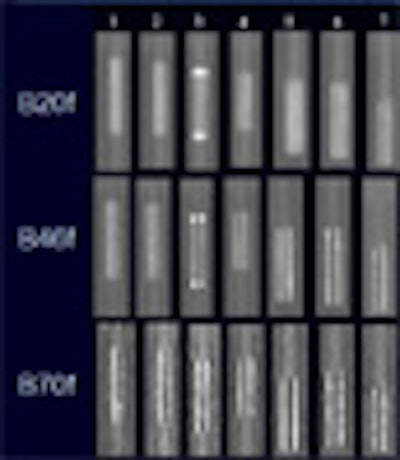
Three different kernels were used for image reconstruction, including the smooth B20f, the sharp B46f, and the very sharp B70f kernel.
"The phantom and the stents were evaluated for artificial lumen narrowing compared with the true lumen of the stent, they were evaluated for the error of intraluminal attenuation values (HU) compared with the true intraluminal attenuation value of 300 HU, and we also looked at artifacts outside the stent, which were measured by the artificial outer diameter widening," Wolf said. "Last but not least, we evaluated the image noise…."
For both artificial lumen narrowing and artifacts outside the stent, the B70f kernel was superior to the B46f kernel, which was superior to the B20f kernel (p < 0.01). Intraluminal attenuation was measured most accurately by the B46f kernel, followed by the B20f kernel, then the B70f kernel (p < 0.01). Image noise was highest with the B70f kernel, followed by the B46f and B20f kernels, with a large overall range of 65 HU (p < 0.01).
 |
| For image reconstruction, three different kernels were used, including the smooth B20f, the sharp B46f, and the very sharp B70f kernel. Axial images and multiplanar reformations were evaluated for artificial lumen narrowing (top chart), error of intraluminal attenuation values (below), artifacts outside the stent (third from top), and image noise (bottom image). For both artificial lumen narrowing and artifacts outside the stent, the B70f kernel was superior to the B46f kernel, which was superior to the B20f kernel (p < 0.01). Intraluminal attenuation was measured most accurately by the B46f kernel, followed by the B20f kernel, then the B70f kernel (p < 0.01). Image noise was highest with the B70f kernel, followed by the B46f and B20f kernels, with a large overall range of 65 HU (p < 0.01). All images courtesy of Dr. Florian Wolf, Medical University of Vienna. |
 |
 |
 |
"Selection of the appropriate reconstruction kernel is crucial for the evaluation of coronary artery stents," Wolf concluded. "Overall, I think the b46f reconstruction kernel can be recommended for the most appropriate reconstruction of coronary artery stents."
 |
| The B46 kernel performed the best overall for stent evaluation. |
Seeing the stent is only part of the issue, cautioned a member of the audience, who asked if the same results could be applied to detecting, for example, intimal hypoplasia. This is a subject for future research, Wolf responded, adding that the ability to see contrast inside the lumen offers clues to other processes if the contrast cannot be visualized.
By Eric Barnes
AuntMinnie.com staff writer
March 11, 2007
Related Reading
Coronary CTA saves money in intermediate-risk individuals, March 10, 2007
U.S. lawmaker seeks data from drug, stent makers, March 7, 2007
Drug-eluting stents reduce subsequent MI frequency and mortality, February 22, 2007
Radiation dose slashed in 64-slice coronary CTA, February 15, 2007
Copyright © 2007 AuntMinnie.com