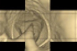
Italian ultrasound vendor Esaote of Genoa is investigating a new ultrasound contrast technology based on the capability of microbubbles to bind themselves to a specific site as an indicator for the presence of cancer.
Esaote collaborated with the University of Genoa, French imaging probe maker Vermon, and Cypriot signal processing specialist SignalGeneriX to develop the ultrasound technique to measure the concentration of microbubbles attached to the targeted indicator to use contrast-enhanced ultrasound for the early detection of cancer.
The three-year research project used in vitro and in vivo tests in mice to demonstrate the technology's potential for the early detection of prostate cancer.
Esaote currently is working to incorporate the technique into its medical imaging equipment by the end of this year. The microbubble agent, however, is unlikely to be available for at least three to five years, due to requirements for clinical trials prior to use in humans.
Related Reading
Biosound Esaote debuts new MyLab units, November 13, 2008
Biosound Esaote to license RCT harmonic patents, January 3, 2008
Biosound Esaote readies ACC introductions, March 20, 2007
Biosound Esaote funds NAPE training, May 3, 2006
Biosound Esaote launches anesthesiology scanner, March 29, 2006
Copyright © 2009 AuntMinnie.com


![Overview of the study design. (A) The fully automated deep learning framework was developed to estimate body composition (BC) (defined as subcutaneous adipose tissue [SAT] in liters; visceral adipose tissue [VAT] in liters; skeletal muscle [SM] in liters; SM fat fraction [SMFF] as a percentage; and intramuscular adipose tissue [IMAT] in deciliters) from MRI. The fully automated framework comprised one model (model 1) to quantify different BC measures (SAT, VAT, SM, SMFF, and IMAT) as three-dimensional (3D) measures from whole-body MRI scans. The second model (model 2) was trained to identify standardized anatomic landmarks along the craniocaudal body axis (z coordinate field), which allowed for subdividing the whole-body measures into different subregions typically examined on clinical routine MRI scans (chest, abdomen, and pelvis). (B) BC was quantified from whole-body MRI in over 66,000 individuals from two large population-based cohort studies, the UK Biobank (UKB) (36,317 individuals) and the German National Cohort (NAKO) (30,291 individuals). Bar graphs show age distribution by sex and cohort. BMI = body mass index. (C) After the performance assessment of the fully automated framework, the change in BC measures, distributions, and profiles across age decades were investigated. Age-, sex-, and height-adjusted body composition reference curves were calculated and made publicly available in a web-based z-score calculator (https://circ-ml.github.io).](https://img.auntminnieeurope.com/mindful/smg/workspaces/default/uploads/2026/05/body-comp.XgAjTfPj1W.jpg?auto=format%2Ccompress&fit=crop&h=112&q=70&w=112)


