Agfa HealthCare announced the integration of Median Technology's Lesion Management Solutions (LMS) computer-aided detection (CAD) software with its IMPAX platform to be used in CT oncology settings.
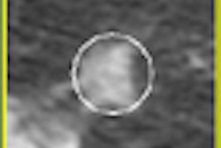
The LMS-Lung and LMS-Liver oncology applications by Median of Sophia Antipolis, France, are designed to assist radiologists to detect, evaluate and follow lesions that have been detected on CT scans. By providing 3D lesion segmentation, measurement calculations, and generated reports, the integration on IMPAX enables desktop access for radiologist review of information, images, and advanced processing across the healthcare enterprise, according the Greenville, SC-based firm. Generated reports then can be transmitted to oncologists and other referring physicians.
Agfa also reported a new installation for its IMPAX RIS/PACS and CR 35-X digitizers, as well as NX workstations, at King Edward VII's Hospital Sister Agnes, a private acute care charitable hospital in London.
Related reading
Agfa shows CR data management software, products at RSNA 2008, November 30, 2008
Agfa lands new Intermountain PACS deal, November 25, 2008
Agfa to debut Impax Scheduling, November 21, 2008
Road to RSNA, CAD, Median Technologies, October 30, 2008
Median nets Spanish contract, March 18, 2008
Copyright © 2008 AuntMinnie.com


![Overview of the study design. (A) The fully automated deep learning framework was developed to estimate body composition (BC) (defined as subcutaneous adipose tissue [SAT] in liters; visceral adipose tissue [VAT] in liters; skeletal muscle [SM] in liters; SM fat fraction [SMFF] as a percentage; and intramuscular adipose tissue [IMAT] in deciliters) from MRI. The fully automated framework comprised one model (model 1) to quantify different BC measures (SAT, VAT, SM, SMFF, and IMAT) as three-dimensional (3D) measures from whole-body MRI scans. The second model (model 2) was trained to identify standardized anatomic landmarks along the craniocaudal body axis (z coordinate field), which allowed for subdividing the whole-body measures into different subregions typically examined on clinical routine MRI scans (chest, abdomen, and pelvis). (B) BC was quantified from whole-body MRI in over 66,000 individuals from two large population-based cohort studies, the UK Biobank (UKB) (36,317 individuals) and the German National Cohort (NAKO) (30,291 individuals). Bar graphs show age distribution by sex and cohort. BMI = body mass index. (C) After the performance assessment of the fully automated framework, the change in BC measures, distributions, and profiles across age decades were investigated. Age-, sex-, and height-adjusted body composition reference curves were calculated and made publicly available in a web-based z-score calculator (https://circ-ml.github.io).](https://img.auntminnieeurope.com/mindful/smg/workspaces/default/uploads/2026/05/body-comp.XgAjTfPj1W.jpg?auto=format%2Ccompress&fit=crop&h=112&q=70&w=112)


