Four experienced pediatric radiologists have shared their knowledge and expertise of orbital anatomy and pathology in new research presented at the annual scientific meeting (ASM) of the Royal Australian and New Zealand College of Radiologists (RANZCR), held in Melbourne from 23 to 25 October.
The authors’ main aims were to provide a simple and concise list of the most frequent orbital pathologies in children; to create a pictorial review with key imaging findings on ultrasound and MRI for general and pediatric radiologists; and to boost understanding of ultrasound findings in pediatric ocular pathology because the exposure to orbital ultrasound is limited.
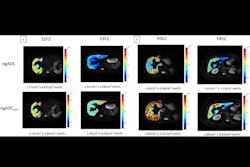
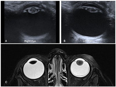 Bilateral congenital cataracts in a 15-day-old infant. Axial ultrasound images of the right lens (A) and left lens (B) demonstrate increased thickness with intralenticular echoes and increased echogenicity of both the anterior and posterior lens walls. (C) Left cataract in a different 2-year-old child with left retinoblastoma. Axial T2-weighted fat-saturated (T2W FS) MRI of the orbits shows globular thickening of the left lens with increased T2 signal compared to the normal right lens, consistent with cataract formation. Additional findings include left retinal detachment and mucosal thickening of the bilateral ethmoid air cells. Attachment of the retinal membrane to the optic disc is not visible in this image. All figures courtesy of Drs. Lasith Ihalagamage, Albert Prat Matifoll, Robert Goetti, and Kristina Prelog and presented at RANZCR's 2025 ASM.
The lack of exposure to orbital ultrasound in many hospitals and the heterogeneous practices in different countries have created significant knowledge gaps in the understanding of technical radiological aspects of ultrasound of the orbits, they explained. Furthermore, most hospitals do not perform ultrasound of the orbits and MRI of the orbits at the same time.
“Given the superficial location of the eye globe and its fluid content, ultrasound is an excellent radiation-free imaging modality to assess the posterior segment of the globe as well as surrounding soft tissues, particularly when ophthalmoscopic evaluation is limited by anterior chamber pathologies,” noted the group, led by Dr. Lasith Ihalagamage, from the Lady Ridgeway Hospital for Children in Colombo, Sri Lanka.
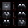
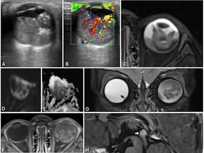 Trilateral retinoblastoma in a 21-month-old child. (A) Axial ultrasound of the left eye shows a solid, heterogeneous mass adherent to the posterior globe wall with echogenic foci suggestive of calcifications. (B) Color Doppler confirms internal vascularity. (C) Axial T2-weighted MRI demonstrates a heterogeneously hypoechoic intraocular lesion with a fluid level. (D-E) The mass is hyperintense on diffusion-weighted imaging and hypointense on the apparent diffusion coefficient (ADC) map, consistent with restricted diffusion. (F) Post-contrast images show heterogeneous enhancement. (G) Coronal T2-weighted image reveals a small nodular soft-tissue elevation in the inferomedial right globe (black arrow), indicating bilateral disease. (H) Sagittal post-contrast T1-weighted image demonstrates an enhancing pineal lesion with associated restricted diffusion (not shown), suggestive of a pineoblastoma, consistent with trilateral retinoblastoma.
The main goal was to gather all the ultrasound and MRI cases in a pediatric hospital during the last 20 years to provide an accessible and concise list of key cases and imaging features to pediatric and general radiologists who are not exposed to ocular ultrasound frequently.
The authors searched the hospital’s ultrasound and MRI electronic databases between January 2004 and December 2024. They collected a total of 584 first diagnostic ultrasound scans and 603 first diagnostic MRI scans of the orbits. The age, gender, clinical symptoms of the patients, and imaging findings were recorded.
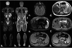
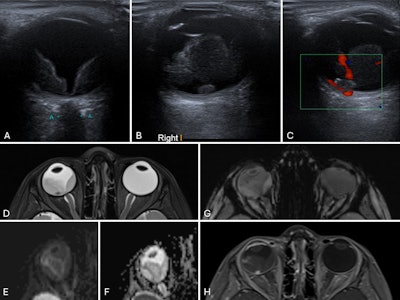 Coats disease in a 3-year-old child. (A) Axial ultrasound of the right eye shows retinal detachment with echogenic subretinal fluid. (B) A small ovoid nodule is seen adjacent to the retina, demonstrating homogeneous echogenicity. (C) Color Doppler imaging shows no internal vascularity within the nodule. (D) Axial T2-weighted MR image of the orbits reveals a slightly small right globe with retinal detachment and subretinal fluid of intermediate signal intensity. The previously identified nodule, located in the foveal region, appears hypointense. (E–F) No evidence of restricted diffusion. (G) Susceptibility-weighted imaging shows no appreciable blooming artifacts to suggest calcification. (H) The lesion demonstrates avid enhancement on post-contrast images. Overall findings are consistent with Coats disease with an enhancing subfoveal nodule.
The most common orbital pathologies detected on ultrasound were hemangioma (92 cases, comprising 15.6% of the total), dermoid/epidermoid (63, 10.8%), optic disc drusen (51, 8.7%), cataract (36, 6.2%), and retinoblastoma (21, 3.6%).
The most common pathologies detected on ultrasound were optic pathway glioma (102 cases, comprising 16.9% of the total), retinoblastoma (55, 9.1%), optic nerve hypoplasia (26, 4.3%), lymphatic malformation (9, 1.49%), and optic neuritis (9, 1.49%).
The researchers focused on the most common intraocular pathologies, including optic disc drusen, papilledema, cataract, retinoblastoma, persistent fetal vasculature, vitreous hemorrhage (nontraumatic), retinal detachment (nontraumatic), Coats disease/exudative retinitis/retinal telangiectasis, trauma-related ocular injuries, and coloboma.
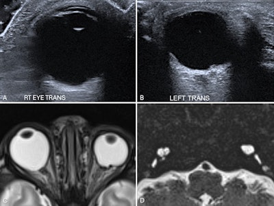 Bilateral coloboma in a 15-day-old neonate with CHARGE syndrome. Ultrasound images of the right eye (A) and left eye (B) show focal defects in the posterior globe with vitreous herniation, suggestive of bilateral colobomas. Axial T2-weighted MR image of the orbits (C) confirms the ultrasound findings. A heavily T2-weighted axial MR image of the petrous temporal bones (D) shows bilaterally dysplastic cochleae and vestibules, with absent semicircular canals -- findings consistent with CHARGE syndrome.
“With proper knowledge of orbital anatomy and pathology, ultrasound serves as a reliable initial imaging modality for evaluating a broad spectrum of orbital pathologies, requiring no special preparation, anesthesia, or sedation,” they wrote.
You can read the full e-poster on the EPOS section for the RANZCR ASM 2025, which is available on the European Society of Radiology's website. The co-authors were Drs. Albert Prat Matifoll, Robert Goetti, and Kristina Prelog, all of the Children's Hospital at Westmead, Sydney Children’s Hospitals Network.


