Radiology plays a key role in characterizing liver lesions and in differential diagnosis, according to a statement issued by the Spanish Society of Medical Radiology (SERAM).
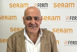
Yet the characterization of newly appearing liver lesions in cancer patients can be a diagnostic challenge, noted specialist Dr. Helena Peris of the Autonomous University of Barcelona. While CT is the gold standard for monitoring cancer patients, liver MRI is considered the most specific test for characterizing liver lesions and is particularly useful when CT findings are atypical for metastasis, Peris noted.
A correct diagnosis requires integrating clinical, analytical, oncological, and imaging information, making a multidisciplinary approach involving oncologists, radiologists, and pathologists essential. Early detection of metastasis has a direct impact on patient survival, as it influences the selection of systemic treatments, targeted therapies, and inclusion in clinical trials, Peris added.
Looking to the future, Peris said that “emerging radiological technologies, as well as integration with AI, is likely to allow radiologists to improve both the detection and characterization of liver lesions.”
The full statement is available on SERAM’s website.