The Spanish Society of Medical Radiology (SERAM) is collaborating with six other scientific societies to develop an AI-supported urology clinical decision-making project called Uro-Oncogu(IA)s.
 The Uro-Oncog(IA)s project team.SERAM
The initiative produced an algorithm that will "reduce time and clinical variability" in the management of urological patients, the society said. SERAM's collaborators include the Spanish Urology Association (AEU), the Foundation for Research in Urology (FIU), the Spanish Society of Pathological Anatomy (SEAP), the Spanish Society of Hospital Pharmacy (SEFH), the Spanish Society of Nuclear Medicine and Molecular Imaging (SEMNIM), and the Spanish Society of Radiation Oncology (SEOR).
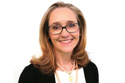
SERAM Secretary General Dr. MaríLuz Parra launched the project in Madrid on 3 July with AEU President Dr. Carmen González.
On behalf of SERAM, the following doctors participated in this initiative:
- Prostate cancer guide: Dr. Joan Carles Vilanova, PhD, of the University of Girona
- Upper urinary tract guide: Dr. Richard Mast of University Hospital Vall d'Hebron in Barcelona
- Muscle-invasive bladder cancer guide: Dr. Eloy Vivas of the University of Malaga
- Non-muscle invasive bladder cancer guide: Dr. Paula Pelechano of the Valencian Institute of Oncology in Valencia
- Kidney cancer guide: Dr. Nicolau Molina of the University of Barcelona.
![A normal mammogram confirmed by three-year radiologic follow-up illustrates reader-marked regions of interest (ROIs) during (A) unaided (round 1) and (B) artificial intelligence (AI)–assisted (round 2) reading. Each colored dot represents an ROI for recall by a human reader. Readers could mark more than one ROI per case, represented by multiple dots of the same color. During AI-assisted reading, the AI system displayed three visible prompts: two with suspicion of malignancy scores of 35% (left mediolateral oblique [L MLO] and craniocaudal [L CC]) and one with a suspicion of malignancy score of 10% (right craniocaudal [R CC]), shown as polygonal overlays. Without AI, six of 10 readers (60%) marked a false-positive ROI. With AI assistance, this fell to two of 10 (20%). R MLO = right mediolateral oblique.](https://img.auntminnieeurope.com/mindful/smg/workspaces/default/uploads/2026/07/2026-07-14-radiology-mammogram-ai-auto-bias.H0bYO8QlWs.jpg?auto=format%2Ccompress&fit=crop&h=100&q=70&w=100)




![A normal mammogram confirmed by three-year radiologic follow-up illustrates reader-marked regions of interest (ROIs) during (A) unaided (round 1) and (B) artificial intelligence (AI)–assisted (round 2) reading. Each colored dot represents an ROI for recall by a human reader. Readers could mark more than one ROI per case, represented by multiple dots of the same color. During AI-assisted reading, the AI system displayed three visible prompts: two with suspicion of malignancy scores of 35% (left mediolateral oblique [L MLO] and craniocaudal [L CC]) and one with a suspicion of malignancy score of 10% (right craniocaudal [R CC]), shown as polygonal overlays. Without AI, six of 10 readers (60%) marked a false-positive ROI. With AI assistance, this fell to two of 10 (20%). R MLO = right mediolateral oblique.](https://img.auntminnieeurope.com/mindful/smg/workspaces/default/uploads/2026/07/2026-07-14-radiology-mammogram-ai-auto-bias.H0bYO8QlWs.jpg?auto=format%2Ccompress&dpr=2&fit=crop&h=167&q=70&w=250)


