Artificial intelligence is helping personalize targeted radionuclide therapy by going beyond receptor expression to analyze tumor radiobiology, optimize individual dosing, and automate complex imaging processes that currently vary dramatically between patients receiving the same treatment.
- Absorbed tumor dose varies dramatically between patients receiving identical activity, sometimes by orders of magnitude, requiring personalized dosing approaches
- AI can reduce imaging requirements, automate tumor segmentation, and adapt treatment cycles based on individual response patterns
- Tumor radiobiology—not just receptor expression—determines treatment response, yet current approaches miss cellular-level effects like crossfire and bystander interactions
- Machine learning models show promise identifying linear energy transfer as a radiation response driver and assessing immune patterns from tissue images
- Prospective data collection and proper data labeling are critical from treatment onset to enable AI-driven treatment refinement and personalization
Find a molecular target, deliver radiation directly to cancer cells, and spare healthy tissue: targeted radionuclide therapy (TRT) promises precision cancer care. But speakers at the RCR meeting in London argued that the field still faces a major personalization challenge.
Absorbed tumor dose can vary dramatically between patients receiving the same activity, sometimes by orders of magnitude, Prof. Jon Wadsley, consultant clinical oncologist at Weston Park Cancer Centre in Sheffield, U.K., highlighted a fundamental limitation.
Some patients may tolerate higher activity and potentially achieve better tumor control, while others may face unnecessary toxicity. In some cases, tumors may absorb too little radiation for additional cycles to provide meaningful benefit.
More personalized approach
The issue is also economic. Wadsley noted that the radionuclide alone for a cycle of lutetium-PSMA therapy costs around £20,000, increasing the need to identify early whether repeated treatment is worthwhile.
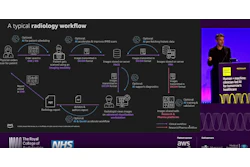
AI approaches could make individualized dosing more practical, he suggested. Current dosimetry requires multiple scans, tumor and organ segmentation, and complex calculations. AI could reduce imaging requirements, automate segmentation, generate dose-volume information, and eventually help adapt treatment cycles according to individual response.
Tumor radiobiology not captured by current approaches
But personalization is not only about measuring dose. Dr. Chris Jones, whose presentation credited CRUK RadNet Cambridge, argued that future models must also explain why tumors respond biologically.
Current selection still relies heavily on whether a tumor expresses the target receptor. Yet some receptor-positive patients respond and others do not, suggesting that tumor radiobiology plays a role not captured by current approaches.
Jones noted that most existing AI radiobiology examples come from external beam radiotherapy, while TRT presents additional complexity. Unlike external beam treatment, TRT delivers radiation that changes across space and time as radiopharmaceuticals distribute through the body, interacting with heterogeneous tumors and differing according to whether beta, alpha, or Auger-electron emitters are used.
He highlighted early AI work relevant to the field, including machine-learning models identifying linear energy transfer as a driver of radiation response, algorithms exploring DNA damage vulnerability, AI-based assessment of immune patterns from H&E-stained tissue images, and models using activity curves to estimate biological effective dose.
Cellular-level radiation effects not fully captured
However, Jones emphasized that major questions remain. Current approaches cannot yet fully capture cellular-level radiation effects, including crossfire and bystander effects between neighboring cells. AI may initially serve as a way to accelerate computationally intensive approaches such as Monte Carlo simulations rather than replace them.
During the panels discussion, session chair Prof. Maria Hawkins of University College London raised a broader concern: once oncology treatments enter practice with fixed regimens, there is often limited incentive to refine who needs more, less, or different treatment. Speakers argued prospective data collection should happen from the beginning rather than years later.
A similar message came from Vanessa Smer-Barreto, PhD, senior postdoctoral fellow at the University of Edinburgh, who presented AI-driven drug discovery work in glioblastoma.
Role of data labeling
Her key point was that data labeling can shape AI performance as much as the model itself. Her group initially classified compounds by whether they reduced cell counts by at least 35%, but this risked detecting general toxicity rather than glioblastoma-specific effects.
Using morphology-informed labeling from cell-painting assays, the researchers aimed to capture richer biological responses. One virtual screen reduced 12,000 candidate compounds to seven selected for laboratory testing, three of which showed activity against glioblastoma cell lines.
A second approach using the molecular language model ChemBERTa-2 narrowed 6 million compounds to roughly 100,000 after applying blood-brain-barrier and structural-diversity filters.
Smer-Barreto emphasized that validation remains essential, including patient-derived organoids and animal models. AI’s immediate role may be less about replacing clinical judgment and more about building the biological understanding needed to personalize future cancer therapy.