The German Röntgen Society (Deutsche Röntgengesellschaft, DRG) has announced that its 2021 doctoral prize has gone to Dr. Quirin Strotzer for his research on deep brain stimulation.
At the 102nd German Radiology Congress, Strotzer, of the University of Regensburg Medical Center, presented new findings that could help to better understand the mechanisms of deep brain stimulation and to improve target point planning for Parkinson's disease and other conditions.
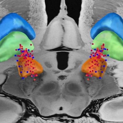
Rendered with Freesurfer -- Template: Edlow 7-tesla MRI of the ex vivo human brain at 100 micron resolution. Images courtesy of Dr. Quirin Strotzer.
Rendered with LEAD-DBS -- Template: Edlow 7-tesla MRI of the ex vivo human brain at 100 micron resolution.Strotzer is a resident in radiology, and he has completed clinical electives and research spells at the University of California, San Diego; the University of British Columbia in Vancouver; and in Helsinki and Moscow.
As part of his doctoral thesis, "Deep brain stimulation: Connectivity profile for bradykinesia alleviation," he published research articles in the Annals of Neurology and Brain Connectivity. He currently conducts research in the field of computational radiology using methods like radiomics and artificial intelligence.




![A normal mammogram confirmed by three-year radiologic follow-up illustrates reader-marked regions of interest (ROIs) during (A) unaided (round 1) and (B) artificial intelligence (AI)–assisted (round 2) reading. Each colored dot represents an ROI for recall by a human reader. Readers could mark more than one ROI per case, represented by multiple dots of the same color. During AI-assisted reading, the AI system displayed three visible prompts: two with suspicion of malignancy scores of 35% (left mediolateral oblique [L MLO] and craniocaudal [L CC]) and one with a suspicion of malignancy score of 10% (right craniocaudal [R CC]), shown as polygonal overlays. Without AI, six of 10 readers (60%) marked a false-positive ROI. With AI assistance, this fell to two of 10 (20%). R MLO = right mediolateral oblique.](https://img.auntminnieeurope.com/mindful/smg/workspaces/default/uploads/2026/07/2026-07-14-radiology-mammogram-ai-auto-bias.H0bYO8QlWs.jpg?auto=format%2Ccompress&fit=crop&h=100&q=70&w=100)


![A normal mammogram confirmed by three-year radiologic follow-up illustrates reader-marked regions of interest (ROIs) during (A) unaided (round 1) and (B) artificial intelligence (AI)–assisted (round 2) reading. Each colored dot represents an ROI for recall by a human reader. Readers could mark more than one ROI per case, represented by multiple dots of the same color. During AI-assisted reading, the AI system displayed three visible prompts: two with suspicion of malignancy scores of 35% (left mediolateral oblique [L MLO] and craniocaudal [L CC]) and one with a suspicion of malignancy score of 10% (right craniocaudal [R CC]), shown as polygonal overlays. Without AI, six of 10 readers (60%) marked a false-positive ROI. With AI assistance, this fell to two of 10 (20%). R MLO = right mediolateral oblique.](https://img.auntminnieeurope.com/mindful/smg/workspaces/default/uploads/2026/07/2026-07-14-radiology-mammogram-ai-auto-bias.H0bYO8QlWs.jpg?auto=format%2Ccompress&fit=crop&h=112&q=70&w=112)