Dear AuntMinnieEurope Member,
In the current era of lockdown and social distancing, webinars are becoming ever more popular. More than 3,000 people logged on recently to hear about Italian radiologists' experiences of COVID-19 cases.
We've posted an article about this European Society of Radiology webinar. It contained a wealth of practical and timely information. Don't miss this report in our CT Community.
PET/CT doesn't instantly spring to mind as a useful modality for COVID-19 patients, but authors from China have presented some results in this area. Their study findings are well worth a close look. Go to the Molecular Imaging Community.
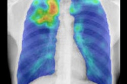
Meanwhile, a group from Radboud University Medical Center in Nijmegen, the Netherlands, has found that artificial intelligence (AI)-based computer-aided detection accurately detects tuberculosis on chest x-rays. The group assessed the performance of the software platform on an independent dataset of over 5,000 chest x-ray images. Head over to the Artificial Intelligence Community.
Is the hype over AI posing a potential risk to patient safety? A team of U.K. and U.S. researchers that includes our columnist Dr. Hugh Harvey believes so. Check out this news report.
MRI has established itself as the primary modality for assessing traumatic brain injuries and strokes. Diffusion-tensor MRI has proved particularly useful in evaluating the brain's white matter and the corpus callosum. Now Swiss researchers have identified several key MRI-based indicators that can help determine how well pediatric patients will recover from a brain injury. Visit the MRI Community.